

JMERC
Owner
Janbazan Medical and Engineering Research Center (JMERC) is the owner of IJWPH.

1.0
Cite Score 2025
SJR: 0.129 / SNIP: 0.140
Volume 18, Issue 2 (2026)
Iran J War Public Health 2026, 18(2): 213-226 |
Back to browse issues page
Article Type:
Subject:




History
Received: 2026/06/1 | Accepted: 2026/07/10 | Published: 2026/07/19
Received: 2026/06/1 | Accepted: 2026/07/10 | Published: 2026/07/19
How to cite this article
Laibi H, Hassani M. L-Glutamine as a Protective Agent Against Carbon Tetrachloride-Induced Liver Injury in Male Mice. Iran J War Public Health 2026; 18 (2) :213-226
URL: http://ijwph.ir/article-1-1785-en.html
URL: http://ijwph.ir/article-1-1785-en.html
Download citation:
BibTeX | RIS | EndNote | Medlars | ProCite | Reference Manager | RefWorks
Send citation to:


Rights and permissions
BibTeX | RIS | EndNote | Medlars | ProCite | Reference Manager | RefWorks
Send citation to:

Authors
H.S. Laibi *1, M.K. Hassani1
1- Department of Biology, College of Science, University of Misan, Maysan, Iraq
Full-Text (HTML) (3 Views)
Introduction
The liver is important for the detoxification and metabolism of many compounds in the human body, whether they are endogenous or exogenous. It is considered the target of many substances’ processing. Its exposure to toxic chemicals may overwhelm its antioxidant defense mechanism and cause hepatocellular damage. Carbon tetrachloride (CCl4) is a toxic chemical molecule that causes liver damage by generating the free-radical trichloromethyl peroxyl radical, which in turn leads to oxidative stress [1]. This hepatocellular injury leads to damage of membrane lipids, enzymes, and DNA [2, 3].
Amino acids (AAs) are among the most vital nutrients for human health. Branched-chain amino acids (BCAAs) are a group of three essential AAs with branched aliphatic side chains: leucine, isoleucine, and valine. BCAAs are involved in energy balance, including gluconeogenesis and lipid metabolism, and have the potential to ameliorate liver problems, in addition to providing a required substrate for protein synthesis [4]. Good nutrition plays a key role in supporting wound healing and tissue regeneration, because the body’s need for nutrients, including AAs, increases under stress conditions, and tissue healing requires cell proliferation. Since some AAs are involved in the synthesis of collagen, the most abundant protein in the body, collagen is essential for maintaining the proper structure and strength of connective tissue, such as bones, skin, cartilage, and blood vessels. Therefore, amino acid support is required for collagen biosynthesis.
Thus, regulating the availability of AAs for collagen biosynthesis is crucial for maintaining tissue integrity, as is the case, for example, during wound healing [5, 6]. On the other hand, exposure of tissues to inflammatory or toxic agents leads to increased levels of free radicals. AAs support cells against oxidative damage by forming glutathione, a tripeptide compound (cysteine, glycine, and glutamic acid) found in relatively high concentrations in many body tissues. It plays a pivotal role in reducing oxidative stress, maintaining redox balance, promoting metabolic detoxification, and regulating the immune system [7].
Glutamine is the most abundant and versatile amino acid in the body, and it is of fundamental importance to intermediary metabolism, interorgan nitrogen exchange via ammonia (NH3) transport between tissues, and pH homeostasis. In almost every cell, glutamine can be used as a substrate for nucleotide synthesis (purines, pyrimidines, and amino sugars), nicotinamide adenine dinucleotide phosphate (NADPH), antioxidants, and many other biosynthetic pathways involved in maintaining cellular integrity and function [8, 9]. Studies indicate that glutamine is a key substrate for providing energy to rapidly dividing cells, particularly in intestinal cells and lymphocytes. The glutamine metabolic pathway begins with deaminase action, producing glutamate and ammonia. Further, glutamate is converted to α-ketoglutarate (α-KG), an intermediate product of the tricarboxylic acid (TCA) cycle, which in turn produces ATP. Consequently, glutamine plays a vital role in increasing cell division, modulating immune defenses, and enhancing protein synthesis efficiency; also, this suggests that, immunologically, glutamine contributes to suppressing inflammatory signaling and protecting cells against oxidative stress and apoptosis [10, 11]. Glutamine has anti-inflammatory properties by influencing a number of inflammatory signaling pathways, including nuclear factor κB (NF-κB) and the signal transducer and activator of transcription (STAT) pathways. In the gastrointestinal tract, glutamine contributes to the integrity of intestinal cells by influencing the expression of tight junction proteins.
This is reflected in the maintenance of the intestinal mucosal barrier and the reduction of permeability [12, 13]. At the organic level, skeletal muscle is the primary source for the synthesis, storage, and release of glutamine into the bloodstream, whereas the intestines, liver, kidneys, and immune system are the main sites of its consumption. Its function in the kidneys is highlighted by its participation in ammonia formation, where its metabolism leads to the production of α-ketoglutarate and bicarbonate ions, which supports regulation of the body’s acid-base balance [14].
This study aimed to investigate the role of L-glutamine against the effect of carbon tetrachloride on the liver of male mice.
Materials and Methods
This experimental study was conducted at the College of Science, University of Misan, on healthy adult mice, aged 12-16 weeks, with an average weight of 25-30g, which were bred at the animal house of the College of Science/University of Misan. The animals were housed in a controlled room under a 12:12 light:dark cycle, randomly separated into plastic cages with hygienic bedding, and fed standard laboratory food. Fifty mice were divided randomly into five groups, each consisting of 10 mice, as follows: G1) untreated group administered with olive oil, which was used as the vehicle; G2) received only CCl4 (1ml/kg body weight): olive oil (1:1) injected intraperitoneally on the 1st and 4th day of every week for 4 weeks; G3) injected with CCl4 (1ml/kg): olive oil (1:1) injected intraperitoneally on the 1st and 4th day of every week for 4 weeks, and then given drinking water containing glutamine (prepared by dissolving 4g of L-glutamine in 1L of drinking water) for 4 weeks; G4) given drinking water containing glutamine and then injected intraperitoneally with CCl4; G5) given only drinking water containing glutamine for 4 weeks [15-17].
Tissue sections were prepared with a rotary microtome (Histoline, Italy) and viewed under a light microscope (Takyo, Japan), and photomicrographs were taken with a digital camera (Canon, Japan) attached to the microscope.
Biochemical methods included separation of blood serum by centrifugation (Hettich, Germany), accurate weighing of samples and materials with a balance (Sartorius, Germany), measurement and adjustment of pH of solutions (Canon, Japan), and storage of samples and reagents at the required temperatures. Serum was also aspirated and transferred using micropipettes (Eppendorf, Germany) to other tubes and spectrophotometrically analyzed.
All animals were anesthetized with chloroform and dissected at 30 days after the initiation of the experiment. Then, blood and liver were collected from the control and treated groups. About 2-3mL of venous blood was collected directly from the heart by puncture using disposable syringes, and the serum was separated, transferred to sterilized tubes, and stored at -20°C for analysis of liver enzymes (alanine transaminase (ALT), aspartate aminotransferase (AST), and alkaline phosphatase (ALP)). The livers from both the experimental and control groups were removed and gathered simultaneously during blood collection, 30 days following the start of the study. The liver samples were sliced into appropriate sizes, rinsed with saline solution, and preserved in a 10% formalin solution. The tissues were then prepared for light microscopy according to the procedure outlined by Luna [18]. Both transverse and longitudinal slices, ranging from 5 to 7µm in thickness, were created and subjected to staining with hematoxylin and eosin (H&E), as well as periodic acid–Schiff (PAS) stains. Finally, the stained samples were analyzed and photographed using a light microscope.
Data analysis was performed using SPSS 23 employing analysis of variance (ANOVA) and least significant difference (LSD).
Findings
Liver samples from control mice exhibited hepatic lobules made up of regular, polygonal hepatocytes with rounded vesicular nuclei and typical cytoplasm. Hepatocytes were arranged into hepatic cords separated by liver sinusoids. Kupffer cells were dispersed among sinusoids, and hepatocytes were predominantly mononucleated, with some binucleated cells present around the central vein. A portal area containing portal tracts and bile ducts was also observed (Figure 1).
Microscopic examination of livers from mice treated intraperitoneally with CCl₄ for 30 days showed prominent histopathological damage, including hepatocyte degeneration and localized cell death around the central vein, vacuolated cytoplasm and shrunken nuclei, increased fatty changes, and disordered hepatic architecture. Portal areas and sinusoids were congested, with inflammatory cell infiltration and degenerative changes in portal structures (Figure 2).
Histopathological examination of livers from the CCl₄ + L-glutamine group showed areas of regeneration. Hepatocytes were arranged in normal sheaths/plates, with clearer cell boundaries and moderately distributed Kupffer cells. Sinusoids were less dilated, the central vein endothelial lining was largely preserved with only mild vacuolation, and periportal architecture was near normal. Mild necrosis and mild inflammatory infiltration were still present, while fatty changes were reduced compared with the CCl₄ group (Figure 3).
Mice livers from the L-glutamine+CCl₄ group showed near-normal histological architecture, with hepatocytes arranged in cords around a central vein and normal cell boundaries. Kupffer cells were normally distributed, sinusoids were less dilated, and there was only mild inflammatory infiltration, with some mildly congested central veins (Figure 4).
The L-glutamine group’s livers showed normal histological architecture, with a central vein surrounded by regular polygonal hepatocytes. Cell boundaries and vesicular nuclei were normal, and hepatocytes were mononucleated and binucleated. Hepatic sinusoids and Kupffer cell distribution were normal, with no evidence of pathological changes (Figure 5).
Control mice stained with PAS to assess glycogen content showed a normal, homogeneous distribution of PAS-positive glycogen granules within hepatocyte cytoplasm, including around the central vein and along cell boundaries (Figure 6).
Microscopic examination of liver sections from the CCl₄ group showed reduced glycogen content within hepatocytes with variable PAS staining, along with mild portal tract wall thickening and mild fatty change (Figure 7).
Microscopic examination of liver sections from the CCl₄ + glutamine-treated group showed a moderate glycogen granule content with a moderate PAS-positive reaction, with granules staining (Figure 8).
Findings from microscopic observation of mice liver sections from glutamine and CCl₄ (G4) showed a strong positive PAS reaction, indicating red or pink granules in hepatocyte cytoplasm (Figure 9).
Microscopic examination of mice liver sections from the glutamine group (G5) showed a strong positive PAS reaction and deposition of coarse glycogen granules (Figure 10).
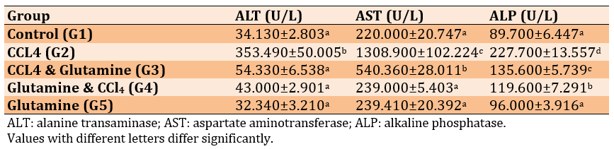
ALT significantly increased in the CCl₄ group (G2) vs. control (p<0.001). AST significantly increased in G2 and G3 vs. control (p<0.001). No significant differences vs. control were found for ALT in G3–G5, for AST in G4–G5, or for ALP in G2–G5 (Table 1).
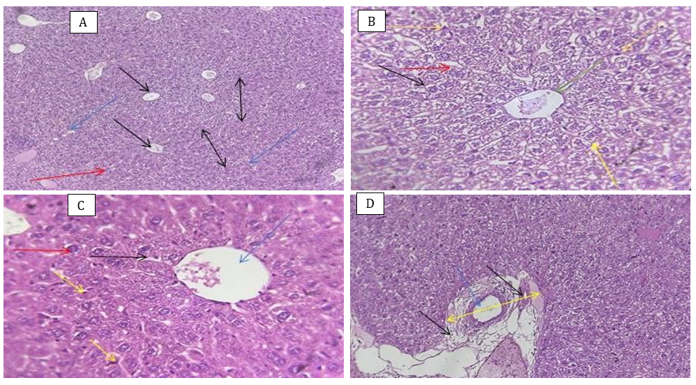
Figure 1. Liver histology of the control (H&E staining).
A) Liver lobules (black double-headed arrow). Each lobule consists of a central vein (single-headed black arrow), hepatocytes arranged as hepatic plates (red arrow), separated from each other by sinusoids (blue arrow). H&E staining (40×).
B) Hepatic cord (blue arrow) separated by smaller sinusoids (red arrow). Hepatocytes (black arrow) around the central vein (green arrow); some hepatocytes are binucleated (yellow arrow), and Kupffer cells (orange arrow) are evident. H&E staining (40×).
C) Hepatocytes around the central vein (blue arrow), each hepatocyte with a single nucleus (red arrow); some others are binucleated (orange arrow). Normal sinusoids (black arrow) and Kupffer cells (yellow arrow) are shown. H&E staining (40×).
D) The portal area (yellow arrow), composed of the portal vein (blue arrow), more connective tissue (black arrow), the hepatic artery (black arrow), and the bile duct (not specified by an arrow color). H&E staining (10×).
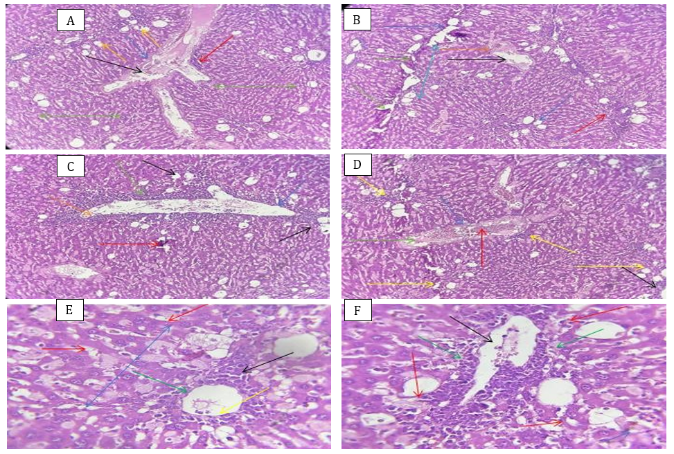
Figure 2. Liver histology of the CCl₄-treated group (H&E staining).
A) Destruction and fibrosis (blue arrow), congested portal tract (black arrow), lymphocytic infiltration (red arrow), disorganized hepatocytes (green arrow), fatty changes (orange arrow), and liver steatosis is shown in the section. H&E staining (10×).
B) Detached hepatocytes (green arrow), with moderate fatty change (blue arrow). The congested portal tract (black arrow) shows accumulation of red blood cells (orange arrow) and infiltration of inflammatory cells (red arrow) extending through degenerated hepatocytes (turquoise arrow). H&E staining (10×).
C) Dilated portal vein (blue arrow), hyperplasia of the lining epithelial layer (orange arrow), inflammatory cells (green arrow), fatty changes such as vesicles (black arrow), and a degenerated region (red arrow). H&E staining (10×).
D) Congested portal tract (blue arrow) with accumulation of red blood cells (red arrow) and inflammatory cells (green arrow). A section in the liver tissue from the CCl4 group showing the congested portal tract (blue arrow), with accumulation of red blood cells (red arrow), and inflammatory cells (green arrow), congested sinusoids (black arrow), and the liver stroma with multiple necrosis foci (yellow arrow). H&E staining (10×).
E) Distortion of hepatocytes (blue arrow) around the central vein (green arrow), degeneration of the endothelial layer (yellow arrow), dilated sinusoids (red arrow), and extensive necrosis (black arrow). H&E staining (40×).
F) Dilated central vein (black arrow) surrounded by thrombosis (green arrow), degeneration of most hepatocytes (red arrow), and cellular fragments (blue arrow). H&E staining (40×).
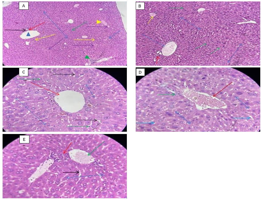
Figure 3. Liver histology of the CCl₄ + glutamine-treated group (H&E staining).
A) Dilated portal tract (blue triangles), bile duct (black arrow), hepatic artery (red arrow), the regenerative area (blue double-headed arrow) obvious, other hepatocytes with clear vesicular nuclei (green arrow), some binucleated (yellow arrow), less dilated sinusoids (blue single-headed arrow), mild necrosis (green arrowhead), mild hemorrhage (yellow arrowhead), Kupffer cells (purple arrow). H&E staining (10x)
B) Dilated portal tract (blue arrow), normal blood vessels (red arrow), mild hemorrhage (yellow arrow), moderate distribution of Kupffer cells (green arrow), irregular septa (purple arrow), and most hepatocytes appeared normal (blue double-headed arrow). H&E staining (10×).
C) A dilated central vein (red arrow), normal regenerated hepatocytes (blue double-headed arrow), normally distributed Kupffer cells (green arrow), and fewer dilated sinusoids (black arrow). H&E staining (40×).
D) A congested central vein (red arrow), mild inflammatory cells (green arrow), normally distributed Kupffer cells (blue single-headed arrow), and most hepatocytes appeared normal (blue double-headed arrow). H&E staining (40×).
E) Normal hepatocytes arranged as cords (blue double-headed arrow) around the congested central vein (green arrow), with mild inflammatory cells (red arrow) obvious, and fewer dilated sinusoids (black arrow). H&E staining (40×).
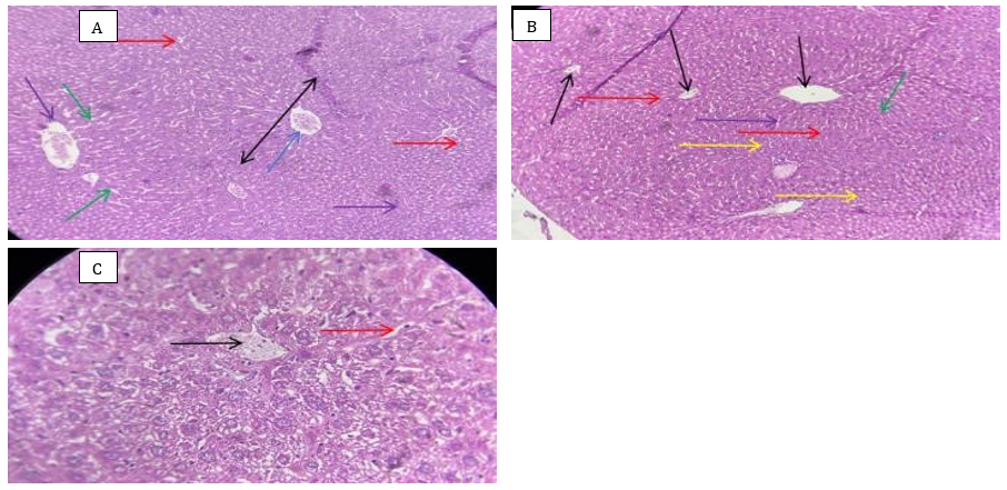
Figure 4. Liver histology of the glutamine + CCl₄-treated group (H&E staining).
A) Normal hepatocytes (black arrow) arranged as cords around a mildly congested central vein (blue arrow), fewer dilated sinusoids (green arrow), and Kupffer cells (red arrow), with mild inflammatory cells (purple arrow). H&E staining (10×).
B) A normal central vein (black arrow), more than one, with normal sinusoids (red arrow), normal distribution of Kupffer cells (yellow arrow), some hepatocytes with a single nucleus (green arrow), other binucleated hepatocytes (purple arrow), and almost normal liver tissue. H&E staining (10×).
C) A mildly congested central vein (black arrow) with normal hepatocytes arranged as cords, fewer dilated sinusoids (red arrow), near-normal architecture with normal cell boundaries. H&E staining (10×).
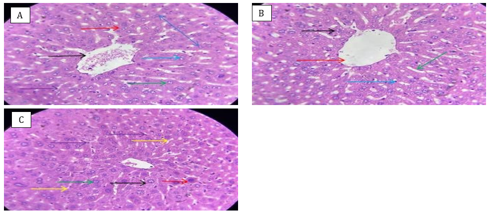
Figure 5. Liver histology of the glutamine-treated group (H&E staining).
A) A central vein (black arrow) surrounded by normal hepatocytes (red arrow), some of which are mononucleated (green arrow), while others are binucleated (blue arrow), arranged in a regular radial pattern (blue double-headed arrow). Hepatic sinusoids appear normal (purple arrow), with no evidence of pathological changes, indicating normal architecture. H&E staining (40×).
B) Normal hepatocytes (black arrow) around the central vein (red arrow), normal sinusoids (green arrow), and Kupffer cells (blue arrow), and hepatic tissue that was normal with no alteration. H&E staining (40×).
C) Polygonal hepatocytes (black arrow) with vesicular nuclei (green arrow), normal cell boundaries (red arrow), normal sinusoids (yellow arrow), Kupffer cells (purple arrow), and hepatic tissue that was normal with no evidence of any changes. H&E staining (40×).
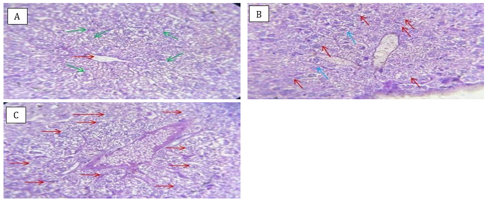
Figure 6. Liver histology of the control (PAS staining).
A) Hepatocytes around the central vein (red arrow) with positive PAS reaction indicating glycogen granule deposition (green arrow). PAS staining (40×).
B) Homogeneous distribution of glycogen granules (red arrow) around faintly stained nuclei (blue arrow). PAS staining (40×).
C) Homogeneous distribution of glycogen granules (red arrow). PAS staining (40×).
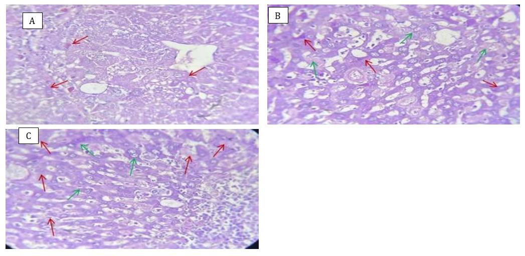
Figure 7. Liver histology of the CCl₄-treated group (PAS stain).
A) A weak PAS reaction indicating mild glycogen content (red arrow) in liver cells. PAS staining (40×).
B) A heterogeneous distribution of glycogen granules, with mild coarse glycogen granules (red arrow) in the hepatocytes’ cytoplasm, and a moderate positive reaction with PAS (green arrow). PAS staining (40×).
C) A heterogeneous distribution of glycogen granules, with mild coarse glycogen granules (red arrow) in the hepatocytes’ cytoplasm, and a moderate positive reaction with PAS (green arrow). PAS staining (40×).
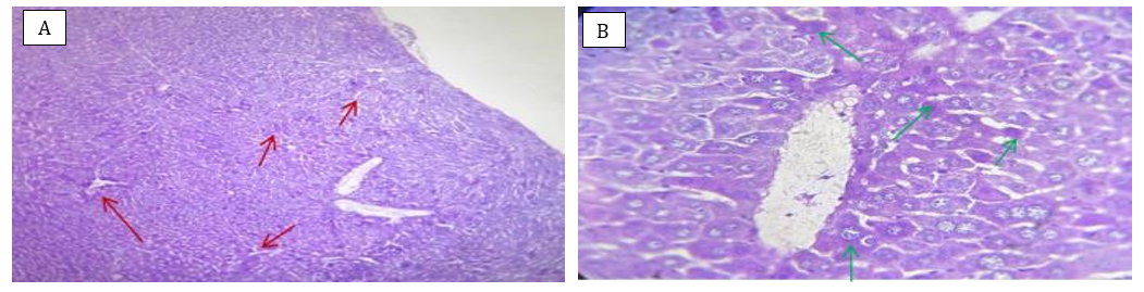
Figure 8. Liver histology of the CCl₄ + glutamine-treated group (PAS staining).
A) A moderate distribution of glycogen granules and a moderate positive reaction with PAS with stained granules (red arrow). PAS staining (10×).
B) A moderate distribution of glycogen granules and a moderate positive reaction with PAS with stained granules (green arrow). PAS staining (40×).
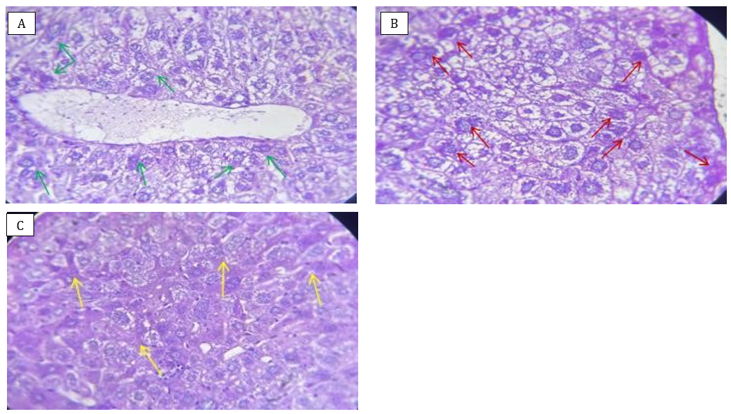
Figure 9. Liver histology of the glutamine + CCl₄-treated group (PAS staining).
A) A strong positive reaction with PAS indicating red or pink granules (green arrow) in the hepatocytes’ cytoplasm. PAS staining (40×).
B) A strong positive reaction with PAS indicating red or pink granules (red arrow) in the hepatocytes’ cytoplasm. PAS staining (40×).
C) A strong positive reaction with PAS indicating red or pink granules (yellow arrow) in the hepatocytes’ cytoplasm. PAS staining (40×).
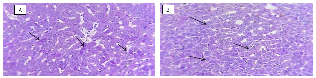
Figure 10. Liver histology of the glutamine-treated group (PAS staining).
A) A strong positive reaction with PAS stain and coarse glycogen granule deposition (black arrow). PAS staining (40×).
B) A strong positive reaction with PAS stain and coarse glycogen granule deposition (black arrow). PAS staining (40×).
Table 1. Mean liver enzyme levels in different groups
Discussion
This study aimed to investigate the role of L-glutamine as a therapeutic and protective agent against the effect of carbon tetrachloride on the liver of male mice. The liver in the control group showed a normal structure, which is consistent with other studies [17, 19, 20], reporting the normal structure of the liver. Liver sections from the mice in the CCl₄ group showed obvious changes, indicating that CCl₄ is considered a toxic agent that is metabolized in the liver and causes liver damage, leading to the production of free radicals and, from there, oxidative stress. This is a common pathological mechanism and contributes to the initiation and progression of liver injury. Oxidative stress also begins to produce inflammatory cytokines, causes hepatocyte necrosis, and stimulates inflammation. These results agree with other research [21, 22], reporting disturbed liver architecture, inflammatory signs, a dilated central vein surrounded by intense cellular infiltration, as well as degenerative changes affecting many hepatocytes. Many cells appeared with dense nuclei and acidophilic cytoplasm. The dilated blood vessels observed can be explained by portal hypertension. Also, the intense cellular infiltration around the central vein can be explained by the induction of inflammation, which is one of the mechanisms of CCl₄-induced liver toxicity.
The liver showed an abnormal structure with a total absence of normal hepatic lobular architecture. Several nodules of varied diameters were separated by thick fibrous septa. In most locations, there was pronounced fatty degeneration of the hepatocytes with scattered necrotic cells. Foci of lobular necrosis with mild inflammation in the portal zone and septa were also seen. The liver also showed significant activity and high degrees of fibrosis. The detected liver damage may be related to the metabolism of CCl₄ into a highly reactive free radical in the liver. This free radical is metabolized by the P450 enzyme system, forming a highly reactive free radical that initiates lipid peroxidation of cytoplasmic membrane phospholipids. This leads to physiological and morphological changes in liver cells, ultimately causing liver damage [23, 24].
CCl₄ alters the retinol pool by impairing CYP4A3 enzymatic activity. This is one of the most significant events that initiates cirrhotic events and upregulates Tgfb, Timp l, and Coll genes, elevates collagen levels, and concomitantly reduces markers associated with antioxidative processes, which are also caused by oxidative stress. In addition, CCl₄-induced oxidative stress increases the level of lipid peroxidation and the production of nitric oxide, leading to an enhanced inflammatory response. Here, the reduction of IL-10 reinforces this imbalance and implies a shift toward a strong proinflammatory environment. In a similar manner, CCl₄ induces an imbalance of hepatic biochemical homeostasis and immune-mediated injury through the upregulation of pro-inflammatory cytokines, consistent with bridging oxidative stress with inflammation [25–27]. Chronic treatment with CCl₄ leads to hepatocellular damage, which in turn activates and induces the proliferation of HSCs, followed by an excessive release of ECM. Eventually, hepatocytes are replaced by excess ECM, which induces scar formation and ultimately leads to fibrosis [28].
Experimental CCl₄ poisoning shows that the glycogen-loaded hepatocyte is much more resistant to injury than one in which fatty infiltration is present, and in which glycogen becomes depleted. That is why chronic alcohol misusers are prone to develop more significant symptoms of CCl₄-induced hepatotoxicity because their liver cells have glycogen depletion and are full [29].
The beneficial effect of L-glutamine was demonstrated in the mice group (G3), where L-glutamine was used as a therapeutic agent. The histological features of the control group indicate partial improvement in the livers of the mice. This partial improvement can be attributed to the fact that when glutamine was administered therapeutically after exposure to CCl₄, liver damage and cellular oxidation had already occurred. Therefore, the role of glutamine was limited to improving and reducing residual oxidative stress and enhancing cellular repair mechanisms.
Our results are in agreement with those of Shrestha et al. [30], who report the beneficial effect of glutamine against CCl₄. They found that glutamine administration significantly reduces collagen deposition and alleviates the degree of fibrosis. Glutamine also reduces the expression of cleaved caspase-3 protein, a reliable marker of apoptosis. The percentage of apoptotic Cahy cells decreases from 4.7% to 2.7% and 2.3% in the 5 mM and 10 mM glutamine-treated groups, respectively. Glutathione depletion has been observed in various experimental models of liver fibrosis. Since glutamine is the primary precursor for the synthesis of the cellular antioxidant glutathione, treatment capable of restoring depleted glutathione content would reverse the process of fibrosis formation.
Several studies have investigated the therapeutic effect of glutamine against other chemical toxins. Schemitt et al. [31] mention the beneficial effect of L-glutamine against thioacetamide. In their study, glutamine reduced lipid peroxidation by 36.36% after 24 hours, 68.97% after 36 hours, and 30.30% after 48 hours. The decrease in TBARS after glutamine administration is also associated with a reduction in necrosis and inflammation scores [32].
Our results contradict those of De Paiva Neto et al. on rats subjected to laparotomy and closure of the hepatic duct and supplemented with 2% glutamine for 21 days. Glutamine treatment does not influence the diagnosis of liver enzymes or histological abnormalities (fibrosis, portal phlebitis, hepatitis, hepatocyte modifications, and bile duct proliferation) in rats with extrahepatic bile duct blockage [33].
Liver sections of the G4 group showed significant improvement, and the liver structure was closer to normal. This can be attributed to the fact that administering glutamine as a protective agent before exposure to CCl₄ was more effective in preventing severe histopathological alterations. It is also due to glutamine’s ability to enhance antioxidants, particularly by increasing the availability of glutathione, which reduces free radicals. In addition, glutamine can suppress the expression of α-SMA and TGF-B in liver tissues and EMT in liver cells. Therefore, it is a possible mechanism involved in preventing liver fibrosis. Our results are consistent with those of Zhang et al. [34], reporting that glutamine is an important substrate for the synthesis of new glutathione, a powerful antioxidant that protects cells from lipid peroxidation. They also showed that glutamine supplementation increases the activity of antioxidant enzymes, including SOD, Gpx, and catalase.
Glutamine supplementation in a high-fat diet inhibited the activation of inflammatory pathways generated by obesity and decreased the priming of immune cell precursors toward a pro-inflammatory phenotype.
In male mice, obesity causes a dramatic shift in structure, with less fat stored in peripheral tissues and in the bones. Glutamine administration alleviates high-fat diet-induced hyperplasia of epididymal white adipose tissue, improved glucose metabolism, and increased bone quality. Additionally, a glutamine-enriched diet enhances cellular metabolism in primary mAT-MSCs and mBMSCs by reestablishing glutamine turnover and reducing inflammation [35].
Liver sections of the G5 group showed normal liver tissue free from changes. This is because glutamine is a naturally occurring amino acid in the body that participates in many metabolic processes and also acts as an antioxidant to maintain the body’s balance.
These results are in agreement with those of De Nóbrega et al. [36]. We did not see any advantageous or disadvantageous effects of glutamine supplementation on biomarkers, including body mass, liver enzyme levels, and histological and functional alterations in the liver. Glutamine is believed to be an endogenous constituent in all tissues and is unlikely to be intrinsically harmful even at elevated concentrations.
Liver sections from control mice stained with PAS for the demonstration of glycogen content showed a normal, homogeneous distribution of fine glycogen granules.
Our results are in agreement with Desouki [37]. Periodic Acid-Schiff (PAS) staining of liver slices from the control animals shows normal glycogen distribution in the central portions and portal regions of the hepatic lobules with increased staining affinity. The glycogen is acidophilic or magenta, and the basement membranes are also red. Arif et al. [38] stained liver slices with PAS dye and examined them under a microscope to establish the presence of glycogen accumulation in the hepatocytes. In their research, the hepatic sections of the control group exhibit a typical and healthy microarchitecture of the liver. Glycogen is observed as a distinct purple-red stain, and cell nuclei are stained light blue. In addition, the central vein, hepatic sinusoids, portal vein, and hepatocytes are all observed to be structurally intact.
Liver from the CCl₄ group showed a mild reduction in glycogen content within hepatocytes. This may be associated with the loss of the liver cells’ functional capacity to store and produce glycogen, and with the detrimental effects of free radicals generated by CCl₄ metabolism. Moreover, lipid alterations due to an imbalance in β-oxidation metabolism lead to fatty acid deoxidation. In addition, CCl₄ promotes an inflammatory response leading to the invasion of immune cells.
Our results are in agreement with Boye et al. [39], who reported that PAS-stained liver sections from model rats show cytoplasmic fat accumulation in centrilobular (hepatocytes surrounding the central vein) and periportal (hepatocytes around the portal triad) areas, together with hepatocytic localized cytoplasmic basophilia. Some cells are positive for PAS, and some cells exhibit decreasing levels of glycogen. In addition, infiltration of glycogen-positive oval cells and well-differentiated hepatocytes with a hyaline appearance is seen in the portal triad region. Additionally, lymphocytic infiltration is observed. The partial reduction in glycogen may be related to enhanced lipogenesis. Furthermore, the significant buildup of vacuolar fat, which pushes the nucleus to the periphery, implies structural and functional failure in the hepatocytes.
Ishikawa et al. [40] and Özerkan et al. [41] report that the livers of CCl₄-exposed animals show PAS-stained hepatocytes surrounding the central vein, although PAS-stained regions corresponding to necrotic areas that had lost glycogen are also observed. This may be explained by the fact that the metabolism of CCl₄ by hepatic microsomal enzymes induces oxidative stress, resulting in damage to cellular lipids and injury to liver cells that have lost their capacity to produce glycogen.
CCl₄ causes hepatic steatosis through a variety of mechanisms, including increased synthesis of fatty acids and triglycerides, synthesis of cholesterol and phospholipids from acetate, and decreased β-oxidation of fatty acids. The researcher also explained that inflammatory lymphocytic infiltration may be related to a greater number of monocytes and lymphocytes, which represents an immune and inflammatory response [42, 43]. Liver sections from the G3 group showed incomplete improvement. There were moderate deposits of glycogen granules with a weakly positive PAS result. This may be due to the fact that, in addition to conserving and reducing glycogen depletion caused by damage, glutamine decreased the severity of liver injury and maintained the metabolic activities of liver cells.
Our results agree with those of Palit et al. [44], reporting an increase in glycogen content, which may be attributed to glutamine’s ability to inhibit gluconeogenesis, decrease hepatic GLUT2, and promote glycolysis and glycogen synthesis. It also improves glucose tolerance and insulin sensitivity by boosting the production of a protein called adiponectin, which is produced by fat cells and helps store glucose in cells.
More study was added by Pitaloka et al. [45], showing that glutamine enhances glycogen synthesis and represses gluconeogenesis. This may be correlated with increased protein expression associated with the PI3K-Akt signaling pathway and decreased expression of G6PC and FOXO1 in hepatocytes.
Liver sections from the G4 group showed a significant improvement and a strong positive PAS result. This might be because glutamine protected liver cells in critical processes and decreased the impacts of stress, thereby reducing cell damage and programmed cell death and enhancing the retention of glycogen reserves in the cell’s intracellular fluid.
Our findings are in agreement with Olaniyi & Olatunji [46]. In their research, pregnant rats received L-glutamine (1g/kg) for 19 days and were subsequently treated with fructose. They reported that ingestion of fructose results in a decrease in liver glycogen quantity and a concomitant decrease in glycogen synthase activity, which may have led to fat accumulation. However, glutamine increases glycogen content, which could be connected to inhibition of the pyruvate dehydrogenase kinase 4, thereby boosting glucose consumption and improving the efficiency of liver glycogen storage.
Another study by Yang et al. [47] on mice found that glutamine enhances glycogen levels, and PAS staining is more apparent than in the alcohol-only group, with poor glycogen levels. This may be related to the fact that glutamine has antioxidant qualities and influences programmed cell death (apoptosis), preventing liver damage and glycogen consumption.
Liver sections from the G5 group showed natural glycogen deposition and strong PAS staining. It is considered a fundamental energy source, and it may also enhance activation of the glycogenolysis pathway by improving liver cell performance and increasing their ability to convert glucose into glycogen.
Our results are in agreement with Miranda et al. [48], in a study on mice given glutamine-enriched food for 20 days; glutamine alone exhibits greater PAS staining than the other groups used.
In G2, there was a significant increase in the levels of liver enzymes ALT, AST, and ALP. This can be explained by the fact that CCl₄ is converted into byproducts that cause harmful effects on the liver, damaging the integrity of its cell membranes; consequently, its enzymes leak into the bloodstream, resulting in the observed increase in enzyme levels.
Our findings are in agreement with the study by Tanvir et al. [49] on male rats, reporting a comparable rise in liver enzymes. This is due to the toxic effects of CCl₄ on the liver, which causes liver damage, cholestasis, and oxidative damage. The findings are also in agreement with those of Madbouly et al. [50], who treated mice with CCl₄ at a dosage of 5ml/kg twice weekly for 3 weeks, reporting a substantial rise in the blood levels of the marker enzymes ALT, AST, and ALP. The rise of these enzymes in CCl₄-treated animals is due to the release of these enzymes into the circulation after rupture of the hepatocyte cell membrane, which enhances cell membrane permeability and favors the migration of extracellular cytoplasmic enzymes [51]. Doma et al. [52] report a substantial rise in the levels of ALT, AST, and ALP enzymes, which is also consistent with earlier studies by Akinloye et al. [53]. This can be explained by the fact that the products of CCl4 metabolism can catalyze lipid peroxidation of the plasma membrane, which can trigger a cascade of reactions, resulting in the deterioration of the integrity of the cell membrane and the subsequent release of enzymes into the extracellular environment [54].
In G3, there was a significant increase in the levels of ALP and AST enzymes. However, no significant differences were observed in ALT compared with the control group. Increased ALT indicates glutamine’s ability to alleviate toxic effects on the liver due to its antioxidant properties, whereas elevated AST is found not only in the liver but also in other organs, such as the heart, kidneys, and muscles. In addition, the ALP enzyme remains elevated because it is closely related to the bile ducts. Therefore, glutamine may repair damage to liver tissue cells, but it does not improve damage to the bile ducts.
Our findings are consistent with those of a study by Shrestha et al. [30] on male mice injected 3 times a week for 10 weeks with 2 different doses of glutamine (0.25 and 0.5g/kg), revealing a reduction and improvement in ALT compared with the CCl₄ group. However, data for additional enzymes are not provided. The reduction in ALT is explained by the fact that glutamine is a primary precursor of the antioxidant glutathione, which decreases free radical-induced oxidation and maintains the integrity of cell membranes. It also lowers TGF-β1 and α-SMA, which are associated with liver fibrosis and, thus, resulting in reduced damage severity. This improves liver health and improves liver enzyme levels. Our findings are somewhat compatible with the following investigations.
In research by Chand [55] on mice, acute liver damage is generated by a single intraperitoneal dose of CCl₄, followed by three successive subcutaneous doses of glutamine. The levels of the aminotransferase enzymes (ALT and AST) are decreased in a dose-dependent manner compared with the CCl₄ group. However, in our research, ALT was closer to normal, while AST and ALP were still elevated. This may be related to the dose and duration of glutamine delivery.
Schemitt et al. [31] report that glutamine reduces the levels of the liver enzymes ALT, AST, and ALP in blood serum. This may be attributed to glutamine’s ability to activate the cellular stress mediator Nrf2, which translocates to the nucleus and binds to DNA via a region known as the antioxidant response element (ARE). This enhances the expression of target genes for cell protection, including antioxidant and detoxification enzymes. Where the gene promoted increased nuclear expression of the Nrf2 protein, while the cytoplasmic expression of the inhibitory protein Keap1 decreased. Glutamine also enhanced the expression of the NQO1 protein and the antioxidant enzyme SOD. Furthermore, glutamine increased GSH levels in liver tissue, leading to reduced inflammation and cell membrane damage, which in turn improved enzyme levels.
Our results contradict the findings of Katoch et al. [56] in male rats with sickle cell anemia, in which L-glutamine is administered at a dose of 10-12mg/mL in their drinking water for 8 weeks. These rats exhibit no improvement in serum markers of liver injury (ALT, AST, and ALP) after L-glutamine treatment.
The researchers found that this is due to advanced disease damage, with ongoing vascular occlusion and Cowper cell hyperplasia, meaning that glutamine is unable to improve the damage to the required extent. Consequently, liver enzyme levels do not improve.
In G4, there was a significant increase in ALP, while no significant differences were observed in the levels of the enzymes ALT and AST compared with the control group. The improvement in liver enzymes ALT and AST indicates the protective efficacy of glutamine in safeguarding liver cells from free radicals, thereby protecting cell membranes from leakage into the bloodstream. While ALP remains elevated, which is more closely associated with the bile ducts, this suggests that glutamine protects liver cells; however, it may not be sufficient to treat cholestasis caused by CCl₄.
Our observations are in agreement with those of Chand [55], generating chronic liver damage by intraperitoneal injection of CCl4 three times weekly for ten weeks, followed by dose-dependent glutamine administration for four weeks after injury. ALP levels are not recorded, although ALT and AST levels drop. The researchers attribute these findings to an effect of glutamine in inhibiting MDA levels and increasing GSH levels. Furthermore, the decreased apoptotic pattern compared with the CCl₄ treatment group implies a protective effect of glutamine on CCl₄-induced hepatocyte damage.
Our findings somewhat accord with the study by Adikwu et al. [57], reporting that glutamine reduces the levels of ALP, AST, and ALP compared with the control group. This differs from our findings, where ALP was reduced while it increased in our present investigation. The researchers credit the reduction in these enzymes to the stabilizing effect of glutamine on the membranes of liver cells, which may have prevented the leaking of these indicators into the circulation. The hepatic oxidative stress generated by BPA might be prevented by glutamine with its antioxidant action (glutathione) [58]. Moreover, glutamine might have repaired liver tissue by stimulating hepatocyte regeneration, development, and repair, leading to improvements in liver enzymes in its cells.
In another study on male mice, Hu et al. [59] found that glutamine lowers plasma levels of liver dysfunction indicators, such as ALT, AST, and LDH compared with the control group. ALP, however, is not affected. The researchers interpret this reduction as a decrease in the production of cleaved caspase-3, thereby reducing the formation of reactive oxygen species in the liver by improving the activity of SOD and GPX as well as GSH levels. Importantly, ALA dramatically decreases the number of phagocytic cells in the liver and inhibited the generation of inflammatory cytokines. All of these elements promote the health of hepatocytes.
In G5, no significant changes or differences were found in the liver enzymes ALT, AST, and ALP compared with the control group. This is attributed to the fact that glutamine has not had a toxic effect at the current dose and duration, thereby maintaining the integrity of liver cell membranes and preventing leakage into the bloodstream. It is also considered a natural amino acid.
The findings of Da Rosa et al.’s study [60] on healthy rats administered L-glutamine at 248mg/kg for 30 days, are comparable with those of a similar study. It is safe for liver tissues and cells, with no notable alterations observed in liver enzymes.
In another study on large rats aged 27 months, the animals are given an intermittent glutamine supplement for 10 months [61]. The study reports no changes in liver enzymes compared with the control, which means there are no positive or negative alterations in hepatotoxicity.
This study provided supportive biochemical and histological evidence that confirms the protective and therapeutic efficacy of glutamine in CCl4-induced hepatotoxicity in mice. Nevertheless, the study has limitations that should be noted. First, although many studies have investigated the molecular effects of glutamine, this study only correlated serum biochemical markers with histopathological changes. It did not measure gene expression for cytokines or cell-death pathways (such as caspases) directly in the liver cells from the mouse samples relevant to this particular experiment. Second, the study was conducted in vivo in mice, and although the data are accurate, further translational studies in humans are needed to enable direct clinical use, taking into account inter-species differences in metabolic pathways. Thus, we suggest the following for future research: complementary studies (histological assessment and gene expression analyses (RT-PCR)) to validate the pathways involved. To further investigate the pharmacokinetics of glutamine and determine the optimal doses to achieve the same histological efficacy observed here before moving to clinical trials.
Conclusion
Glutamine both protects against and therapeutically attenuates CCl₄-induced liver injury, with a stronger protective effect when administered as pretreatment and corresponding improvements in liver enzyme biomarkers.
Acknowledgments: The authors would like to express their sincere gratitude and appreciation to the Department of Biology, College of Science, University of Misan, for providing the necessary laboratory facilities and administrative support to complete this research.
Ethical Permissions: All experimental procedures involving animals were approved by the Research Ethics Committee of the College of Science, University of Misan, Iraq.
Conflicts of Interests: The authors declared no conflicts of interest.
Authors' Contribution: Laibi HS (First Author), Introduction Writer/Methodologist/Main Researcher/Discussion Writer/Statistical Analyst (70%); Hassani MK (Second Author), Methodologist/Assistant Researcher/Statistical Analyst (30%)
Funding/Support: The authors received no financial support or funding for the research, authorship, and/or publication of this article. This research was self-funded.
The liver is important for the detoxification and metabolism of many compounds in the human body, whether they are endogenous or exogenous. It is considered the target of many substances’ processing. Its exposure to toxic chemicals may overwhelm its antioxidant defense mechanism and cause hepatocellular damage. Carbon tetrachloride (CCl4) is a toxic chemical molecule that causes liver damage by generating the free-radical trichloromethyl peroxyl radical, which in turn leads to oxidative stress [1]. This hepatocellular injury leads to damage of membrane lipids, enzymes, and DNA [2, 3].
Amino acids (AAs) are among the most vital nutrients for human health. Branched-chain amino acids (BCAAs) are a group of three essential AAs with branched aliphatic side chains: leucine, isoleucine, and valine. BCAAs are involved in energy balance, including gluconeogenesis and lipid metabolism, and have the potential to ameliorate liver problems, in addition to providing a required substrate for protein synthesis [4]. Good nutrition plays a key role in supporting wound healing and tissue regeneration, because the body’s need for nutrients, including AAs, increases under stress conditions, and tissue healing requires cell proliferation. Since some AAs are involved in the synthesis of collagen, the most abundant protein in the body, collagen is essential for maintaining the proper structure and strength of connective tissue, such as bones, skin, cartilage, and blood vessels. Therefore, amino acid support is required for collagen biosynthesis.
Thus, regulating the availability of AAs for collagen biosynthesis is crucial for maintaining tissue integrity, as is the case, for example, during wound healing [5, 6]. On the other hand, exposure of tissues to inflammatory or toxic agents leads to increased levels of free radicals. AAs support cells against oxidative damage by forming glutathione, a tripeptide compound (cysteine, glycine, and glutamic acid) found in relatively high concentrations in many body tissues. It plays a pivotal role in reducing oxidative stress, maintaining redox balance, promoting metabolic detoxification, and regulating the immune system [7].
Glutamine is the most abundant and versatile amino acid in the body, and it is of fundamental importance to intermediary metabolism, interorgan nitrogen exchange via ammonia (NH3) transport between tissues, and pH homeostasis. In almost every cell, glutamine can be used as a substrate for nucleotide synthesis (purines, pyrimidines, and amino sugars), nicotinamide adenine dinucleotide phosphate (NADPH), antioxidants, and many other biosynthetic pathways involved in maintaining cellular integrity and function [8, 9]. Studies indicate that glutamine is a key substrate for providing energy to rapidly dividing cells, particularly in intestinal cells and lymphocytes. The glutamine metabolic pathway begins with deaminase action, producing glutamate and ammonia. Further, glutamate is converted to α-ketoglutarate (α-KG), an intermediate product of the tricarboxylic acid (TCA) cycle, which in turn produces ATP. Consequently, glutamine plays a vital role in increasing cell division, modulating immune defenses, and enhancing protein synthesis efficiency; also, this suggests that, immunologically, glutamine contributes to suppressing inflammatory signaling and protecting cells against oxidative stress and apoptosis [10, 11]. Glutamine has anti-inflammatory properties by influencing a number of inflammatory signaling pathways, including nuclear factor κB (NF-κB) and the signal transducer and activator of transcription (STAT) pathways. In the gastrointestinal tract, glutamine contributes to the integrity of intestinal cells by influencing the expression of tight junction proteins.
This is reflected in the maintenance of the intestinal mucosal barrier and the reduction of permeability [12, 13]. At the organic level, skeletal muscle is the primary source for the synthesis, storage, and release of glutamine into the bloodstream, whereas the intestines, liver, kidneys, and immune system are the main sites of its consumption. Its function in the kidneys is highlighted by its participation in ammonia formation, where its metabolism leads to the production of α-ketoglutarate and bicarbonate ions, which supports regulation of the body’s acid-base balance [14].
This study aimed to investigate the role of L-glutamine against the effect of carbon tetrachloride on the liver of male mice.
Materials and Methods
This experimental study was conducted at the College of Science, University of Misan, on healthy adult mice, aged 12-16 weeks, with an average weight of 25-30g, which were bred at the animal house of the College of Science/University of Misan. The animals were housed in a controlled room under a 12:12 light:dark cycle, randomly separated into plastic cages with hygienic bedding, and fed standard laboratory food. Fifty mice were divided randomly into five groups, each consisting of 10 mice, as follows: G1) untreated group administered with olive oil, which was used as the vehicle; G2) received only CCl4 (1ml/kg body weight): olive oil (1:1) injected intraperitoneally on the 1st and 4th day of every week for 4 weeks; G3) injected with CCl4 (1ml/kg): olive oil (1:1) injected intraperitoneally on the 1st and 4th day of every week for 4 weeks, and then given drinking water containing glutamine (prepared by dissolving 4g of L-glutamine in 1L of drinking water) for 4 weeks; G4) given drinking water containing glutamine and then injected intraperitoneally with CCl4; G5) given only drinking water containing glutamine for 4 weeks [15-17].
Tissue sections were prepared with a rotary microtome (Histoline, Italy) and viewed under a light microscope (Takyo, Japan), and photomicrographs were taken with a digital camera (Canon, Japan) attached to the microscope.
Biochemical methods included separation of blood serum by centrifugation (Hettich, Germany), accurate weighing of samples and materials with a balance (Sartorius, Germany), measurement and adjustment of pH of solutions (Canon, Japan), and storage of samples and reagents at the required temperatures. Serum was also aspirated and transferred using micropipettes (Eppendorf, Germany) to other tubes and spectrophotometrically analyzed.
All animals were anesthetized with chloroform and dissected at 30 days after the initiation of the experiment. Then, blood and liver were collected from the control and treated groups. About 2-3mL of venous blood was collected directly from the heart by puncture using disposable syringes, and the serum was separated, transferred to sterilized tubes, and stored at -20°C for analysis of liver enzymes (alanine transaminase (ALT), aspartate aminotransferase (AST), and alkaline phosphatase (ALP)). The livers from both the experimental and control groups were removed and gathered simultaneously during blood collection, 30 days following the start of the study. The liver samples were sliced into appropriate sizes, rinsed with saline solution, and preserved in a 10% formalin solution. The tissues were then prepared for light microscopy according to the procedure outlined by Luna [18]. Both transverse and longitudinal slices, ranging from 5 to 7µm in thickness, were created and subjected to staining with hematoxylin and eosin (H&E), as well as periodic acid–Schiff (PAS) stains. Finally, the stained samples were analyzed and photographed using a light microscope.
Data analysis was performed using SPSS 23 employing analysis of variance (ANOVA) and least significant difference (LSD).
Findings
Liver samples from control mice exhibited hepatic lobules made up of regular, polygonal hepatocytes with rounded vesicular nuclei and typical cytoplasm. Hepatocytes were arranged into hepatic cords separated by liver sinusoids. Kupffer cells were dispersed among sinusoids, and hepatocytes were predominantly mononucleated, with some binucleated cells present around the central vein. A portal area containing portal tracts and bile ducts was also observed (Figure 1).
Microscopic examination of livers from mice treated intraperitoneally with CCl₄ for 30 days showed prominent histopathological damage, including hepatocyte degeneration and localized cell death around the central vein, vacuolated cytoplasm and shrunken nuclei, increased fatty changes, and disordered hepatic architecture. Portal areas and sinusoids were congested, with inflammatory cell infiltration and degenerative changes in portal structures (Figure 2).
Histopathological examination of livers from the CCl₄ + L-glutamine group showed areas of regeneration. Hepatocytes were arranged in normal sheaths/plates, with clearer cell boundaries and moderately distributed Kupffer cells. Sinusoids were less dilated, the central vein endothelial lining was largely preserved with only mild vacuolation, and periportal architecture was near normal. Mild necrosis and mild inflammatory infiltration were still present, while fatty changes were reduced compared with the CCl₄ group (Figure 3).
Mice livers from the L-glutamine+CCl₄ group showed near-normal histological architecture, with hepatocytes arranged in cords around a central vein and normal cell boundaries. Kupffer cells were normally distributed, sinusoids were less dilated, and there was only mild inflammatory infiltration, with some mildly congested central veins (Figure 4).
The L-glutamine group’s livers showed normal histological architecture, with a central vein surrounded by regular polygonal hepatocytes. Cell boundaries and vesicular nuclei were normal, and hepatocytes were mononucleated and binucleated. Hepatic sinusoids and Kupffer cell distribution were normal, with no evidence of pathological changes (Figure 5).
Control mice stained with PAS to assess glycogen content showed a normal, homogeneous distribution of PAS-positive glycogen granules within hepatocyte cytoplasm, including around the central vein and along cell boundaries (Figure 6).
Microscopic examination of liver sections from the CCl₄ group showed reduced glycogen content within hepatocytes with variable PAS staining, along with mild portal tract wall thickening and mild fatty change (Figure 7).
Microscopic examination of liver sections from the CCl₄ + glutamine-treated group showed a moderate glycogen granule content with a moderate PAS-positive reaction, with granules staining (Figure 8).
Findings from microscopic observation of mice liver sections from glutamine and CCl₄ (G4) showed a strong positive PAS reaction, indicating red or pink granules in hepatocyte cytoplasm (Figure 9).
Microscopic examination of mice liver sections from the glutamine group (G5) showed a strong positive PAS reaction and deposition of coarse glycogen granules (Figure 10).
ALT significantly increased in the CCl₄ group (G2) vs. control (p<0.001). AST significantly increased in G2 and G3 vs. control (p<0.001). No significant differences vs. control were found for ALT in G3–G5, for AST in G4–G5, or for ALP in G2–G5 (Table 1).
Figure 1. Liver histology of the control (H&E staining).
A) Liver lobules (black double-headed arrow). Each lobule consists of a central vein (single-headed black arrow), hepatocytes arranged as hepatic plates (red arrow), separated from each other by sinusoids (blue arrow). H&E staining (40×).
B) Hepatic cord (blue arrow) separated by smaller sinusoids (red arrow). Hepatocytes (black arrow) around the central vein (green arrow); some hepatocytes are binucleated (yellow arrow), and Kupffer cells (orange arrow) are evident. H&E staining (40×).
C) Hepatocytes around the central vein (blue arrow), each hepatocyte with a single nucleus (red arrow); some others are binucleated (orange arrow). Normal sinusoids (black arrow) and Kupffer cells (yellow arrow) are shown. H&E staining (40×).
D) The portal area (yellow arrow), composed of the portal vein (blue arrow), more connective tissue (black arrow), the hepatic artery (black arrow), and the bile duct (not specified by an arrow color). H&E staining (10×).
Figure 2. Liver histology of the CCl₄-treated group (H&E staining).
A) Destruction and fibrosis (blue arrow), congested portal tract (black arrow), lymphocytic infiltration (red arrow), disorganized hepatocytes (green arrow), fatty changes (orange arrow), and liver steatosis is shown in the section. H&E staining (10×).
B) Detached hepatocytes (green arrow), with moderate fatty change (blue arrow). The congested portal tract (black arrow) shows accumulation of red blood cells (orange arrow) and infiltration of inflammatory cells (red arrow) extending through degenerated hepatocytes (turquoise arrow). H&E staining (10×).
C) Dilated portal vein (blue arrow), hyperplasia of the lining epithelial layer (orange arrow), inflammatory cells (green arrow), fatty changes such as vesicles (black arrow), and a degenerated region (red arrow). H&E staining (10×).
D) Congested portal tract (blue arrow) with accumulation of red blood cells (red arrow) and inflammatory cells (green arrow). A section in the liver tissue from the CCl4 group showing the congested portal tract (blue arrow), with accumulation of red blood cells (red arrow), and inflammatory cells (green arrow), congested sinusoids (black arrow), and the liver stroma with multiple necrosis foci (yellow arrow). H&E staining (10×).
E) Distortion of hepatocytes (blue arrow) around the central vein (green arrow), degeneration of the endothelial layer (yellow arrow), dilated sinusoids (red arrow), and extensive necrosis (black arrow). H&E staining (40×).
F) Dilated central vein (black arrow) surrounded by thrombosis (green arrow), degeneration of most hepatocytes (red arrow), and cellular fragments (blue arrow). H&E staining (40×).
Figure 3. Liver histology of the CCl₄ + glutamine-treated group (H&E staining).
A) Dilated portal tract (blue triangles), bile duct (black arrow), hepatic artery (red arrow), the regenerative area (blue double-headed arrow) obvious, other hepatocytes with clear vesicular nuclei (green arrow), some binucleated (yellow arrow), less dilated sinusoids (blue single-headed arrow), mild necrosis (green arrowhead), mild hemorrhage (yellow arrowhead), Kupffer cells (purple arrow). H&E staining (10x)
B) Dilated portal tract (blue arrow), normal blood vessels (red arrow), mild hemorrhage (yellow arrow), moderate distribution of Kupffer cells (green arrow), irregular septa (purple arrow), and most hepatocytes appeared normal (blue double-headed arrow). H&E staining (10×).
C) A dilated central vein (red arrow), normal regenerated hepatocytes (blue double-headed arrow), normally distributed Kupffer cells (green arrow), and fewer dilated sinusoids (black arrow). H&E staining (40×).
D) A congested central vein (red arrow), mild inflammatory cells (green arrow), normally distributed Kupffer cells (blue single-headed arrow), and most hepatocytes appeared normal (blue double-headed arrow). H&E staining (40×).
E) Normal hepatocytes arranged as cords (blue double-headed arrow) around the congested central vein (green arrow), with mild inflammatory cells (red arrow) obvious, and fewer dilated sinusoids (black arrow). H&E staining (40×).
Figure 4. Liver histology of the glutamine + CCl₄-treated group (H&E staining).
A) Normal hepatocytes (black arrow) arranged as cords around a mildly congested central vein (blue arrow), fewer dilated sinusoids (green arrow), and Kupffer cells (red arrow), with mild inflammatory cells (purple arrow). H&E staining (10×).
B) A normal central vein (black arrow), more than one, with normal sinusoids (red arrow), normal distribution of Kupffer cells (yellow arrow), some hepatocytes with a single nucleus (green arrow), other binucleated hepatocytes (purple arrow), and almost normal liver tissue. H&E staining (10×).
C) A mildly congested central vein (black arrow) with normal hepatocytes arranged as cords, fewer dilated sinusoids (red arrow), near-normal architecture with normal cell boundaries. H&E staining (10×).
Figure 5. Liver histology of the glutamine-treated group (H&E staining).
A) A central vein (black arrow) surrounded by normal hepatocytes (red arrow), some of which are mononucleated (green arrow), while others are binucleated (blue arrow), arranged in a regular radial pattern (blue double-headed arrow). Hepatic sinusoids appear normal (purple arrow), with no evidence of pathological changes, indicating normal architecture. H&E staining (40×).
B) Normal hepatocytes (black arrow) around the central vein (red arrow), normal sinusoids (green arrow), and Kupffer cells (blue arrow), and hepatic tissue that was normal with no alteration. H&E staining (40×).
C) Polygonal hepatocytes (black arrow) with vesicular nuclei (green arrow), normal cell boundaries (red arrow), normal sinusoids (yellow arrow), Kupffer cells (purple arrow), and hepatic tissue that was normal with no evidence of any changes. H&E staining (40×).
Figure 6. Liver histology of the control (PAS staining).
A) Hepatocytes around the central vein (red arrow) with positive PAS reaction indicating glycogen granule deposition (green arrow). PAS staining (40×).
B) Homogeneous distribution of glycogen granules (red arrow) around faintly stained nuclei (blue arrow). PAS staining (40×).
C) Homogeneous distribution of glycogen granules (red arrow). PAS staining (40×).
Figure 7. Liver histology of the CCl₄-treated group (PAS stain).
A) A weak PAS reaction indicating mild glycogen content (red arrow) in liver cells. PAS staining (40×).
B) A heterogeneous distribution of glycogen granules, with mild coarse glycogen granules (red arrow) in the hepatocytes’ cytoplasm, and a moderate positive reaction with PAS (green arrow). PAS staining (40×).
C) A heterogeneous distribution of glycogen granules, with mild coarse glycogen granules (red arrow) in the hepatocytes’ cytoplasm, and a moderate positive reaction with PAS (green arrow). PAS staining (40×).
Figure 8. Liver histology of the CCl₄ + glutamine-treated group (PAS staining).
A) A moderate distribution of glycogen granules and a moderate positive reaction with PAS with stained granules (red arrow). PAS staining (10×).
B) A moderate distribution of glycogen granules and a moderate positive reaction with PAS with stained granules (green arrow). PAS staining (40×).
Figure 9. Liver histology of the glutamine + CCl₄-treated group (PAS staining).
A) A strong positive reaction with PAS indicating red or pink granules (green arrow) in the hepatocytes’ cytoplasm. PAS staining (40×).
B) A strong positive reaction with PAS indicating red or pink granules (red arrow) in the hepatocytes’ cytoplasm. PAS staining (40×).
C) A strong positive reaction with PAS indicating red or pink granules (yellow arrow) in the hepatocytes’ cytoplasm. PAS staining (40×).
Figure 10. Liver histology of the glutamine-treated group (PAS staining).
A) A strong positive reaction with PAS stain and coarse glycogen granule deposition (black arrow). PAS staining (40×).
B) A strong positive reaction with PAS stain and coarse glycogen granule deposition (black arrow). PAS staining (40×).
Table 1. Mean liver enzyme levels in different groups
Discussion
This study aimed to investigate the role of L-glutamine as a therapeutic and protective agent against the effect of carbon tetrachloride on the liver of male mice. The liver in the control group showed a normal structure, which is consistent with other studies [17, 19, 20], reporting the normal structure of the liver. Liver sections from the mice in the CCl₄ group showed obvious changes, indicating that CCl₄ is considered a toxic agent that is metabolized in the liver and causes liver damage, leading to the production of free radicals and, from there, oxidative stress. This is a common pathological mechanism and contributes to the initiation and progression of liver injury. Oxidative stress also begins to produce inflammatory cytokines, causes hepatocyte necrosis, and stimulates inflammation. These results agree with other research [21, 22], reporting disturbed liver architecture, inflammatory signs, a dilated central vein surrounded by intense cellular infiltration, as well as degenerative changes affecting many hepatocytes. Many cells appeared with dense nuclei and acidophilic cytoplasm. The dilated blood vessels observed can be explained by portal hypertension. Also, the intense cellular infiltration around the central vein can be explained by the induction of inflammation, which is one of the mechanisms of CCl₄-induced liver toxicity.
The liver showed an abnormal structure with a total absence of normal hepatic lobular architecture. Several nodules of varied diameters were separated by thick fibrous septa. In most locations, there was pronounced fatty degeneration of the hepatocytes with scattered necrotic cells. Foci of lobular necrosis with mild inflammation in the portal zone and septa were also seen. The liver also showed significant activity and high degrees of fibrosis. The detected liver damage may be related to the metabolism of CCl₄ into a highly reactive free radical in the liver. This free radical is metabolized by the P450 enzyme system, forming a highly reactive free radical that initiates lipid peroxidation of cytoplasmic membrane phospholipids. This leads to physiological and morphological changes in liver cells, ultimately causing liver damage [23, 24].
CCl₄ alters the retinol pool by impairing CYP4A3 enzymatic activity. This is one of the most significant events that initiates cirrhotic events and upregulates Tgfb, Timp l, and Coll genes, elevates collagen levels, and concomitantly reduces markers associated with antioxidative processes, which are also caused by oxidative stress. In addition, CCl₄-induced oxidative stress increases the level of lipid peroxidation and the production of nitric oxide, leading to an enhanced inflammatory response. Here, the reduction of IL-10 reinforces this imbalance and implies a shift toward a strong proinflammatory environment. In a similar manner, CCl₄ induces an imbalance of hepatic biochemical homeostasis and immune-mediated injury through the upregulation of pro-inflammatory cytokines, consistent with bridging oxidative stress with inflammation [25–27]. Chronic treatment with CCl₄ leads to hepatocellular damage, which in turn activates and induces the proliferation of HSCs, followed by an excessive release of ECM. Eventually, hepatocytes are replaced by excess ECM, which induces scar formation and ultimately leads to fibrosis [28].
Experimental CCl₄ poisoning shows that the glycogen-loaded hepatocyte is much more resistant to injury than one in which fatty infiltration is present, and in which glycogen becomes depleted. That is why chronic alcohol misusers are prone to develop more significant symptoms of CCl₄-induced hepatotoxicity because their liver cells have glycogen depletion and are full [29].
The beneficial effect of L-glutamine was demonstrated in the mice group (G3), where L-glutamine was used as a therapeutic agent. The histological features of the control group indicate partial improvement in the livers of the mice. This partial improvement can be attributed to the fact that when glutamine was administered therapeutically after exposure to CCl₄, liver damage and cellular oxidation had already occurred. Therefore, the role of glutamine was limited to improving and reducing residual oxidative stress and enhancing cellular repair mechanisms.
Our results are in agreement with those of Shrestha et al. [30], who report the beneficial effect of glutamine against CCl₄. They found that glutamine administration significantly reduces collagen deposition and alleviates the degree of fibrosis. Glutamine also reduces the expression of cleaved caspase-3 protein, a reliable marker of apoptosis. The percentage of apoptotic Cahy cells decreases from 4.7% to 2.7% and 2.3% in the 5 mM and 10 mM glutamine-treated groups, respectively. Glutathione depletion has been observed in various experimental models of liver fibrosis. Since glutamine is the primary precursor for the synthesis of the cellular antioxidant glutathione, treatment capable of restoring depleted glutathione content would reverse the process of fibrosis formation.
Several studies have investigated the therapeutic effect of glutamine against other chemical toxins. Schemitt et al. [31] mention the beneficial effect of L-glutamine against thioacetamide. In their study, glutamine reduced lipid peroxidation by 36.36% after 24 hours, 68.97% after 36 hours, and 30.30% after 48 hours. The decrease in TBARS after glutamine administration is also associated with a reduction in necrosis and inflammation scores [32].
Our results contradict those of De Paiva Neto et al. on rats subjected to laparotomy and closure of the hepatic duct and supplemented with 2% glutamine for 21 days. Glutamine treatment does not influence the diagnosis of liver enzymes or histological abnormalities (fibrosis, portal phlebitis, hepatitis, hepatocyte modifications, and bile duct proliferation) in rats with extrahepatic bile duct blockage [33].
Liver sections of the G4 group showed significant improvement, and the liver structure was closer to normal. This can be attributed to the fact that administering glutamine as a protective agent before exposure to CCl₄ was more effective in preventing severe histopathological alterations. It is also due to glutamine’s ability to enhance antioxidants, particularly by increasing the availability of glutathione, which reduces free radicals. In addition, glutamine can suppress the expression of α-SMA and TGF-B in liver tissues and EMT in liver cells. Therefore, it is a possible mechanism involved in preventing liver fibrosis. Our results are consistent with those of Zhang et al. [34], reporting that glutamine is an important substrate for the synthesis of new glutathione, a powerful antioxidant that protects cells from lipid peroxidation. They also showed that glutamine supplementation increases the activity of antioxidant enzymes, including SOD, Gpx, and catalase.
Glutamine supplementation in a high-fat diet inhibited the activation of inflammatory pathways generated by obesity and decreased the priming of immune cell precursors toward a pro-inflammatory phenotype.
In male mice, obesity causes a dramatic shift in structure, with less fat stored in peripheral tissues and in the bones. Glutamine administration alleviates high-fat diet-induced hyperplasia of epididymal white adipose tissue, improved glucose metabolism, and increased bone quality. Additionally, a glutamine-enriched diet enhances cellular metabolism in primary mAT-MSCs and mBMSCs by reestablishing glutamine turnover and reducing inflammation [35].
Liver sections of the G5 group showed normal liver tissue free from changes. This is because glutamine is a naturally occurring amino acid in the body that participates in many metabolic processes and also acts as an antioxidant to maintain the body’s balance.
These results are in agreement with those of De Nóbrega et al. [36]. We did not see any advantageous or disadvantageous effects of glutamine supplementation on biomarkers, including body mass, liver enzyme levels, and histological and functional alterations in the liver. Glutamine is believed to be an endogenous constituent in all tissues and is unlikely to be intrinsically harmful even at elevated concentrations.
Liver sections from control mice stained with PAS for the demonstration of glycogen content showed a normal, homogeneous distribution of fine glycogen granules.
Our results are in agreement with Desouki [37]. Periodic Acid-Schiff (PAS) staining of liver slices from the control animals shows normal glycogen distribution in the central portions and portal regions of the hepatic lobules with increased staining affinity. The glycogen is acidophilic or magenta, and the basement membranes are also red. Arif et al. [38] stained liver slices with PAS dye and examined them under a microscope to establish the presence of glycogen accumulation in the hepatocytes. In their research, the hepatic sections of the control group exhibit a typical and healthy microarchitecture of the liver. Glycogen is observed as a distinct purple-red stain, and cell nuclei are stained light blue. In addition, the central vein, hepatic sinusoids, portal vein, and hepatocytes are all observed to be structurally intact.
Liver from the CCl₄ group showed a mild reduction in glycogen content within hepatocytes. This may be associated with the loss of the liver cells’ functional capacity to store and produce glycogen, and with the detrimental effects of free radicals generated by CCl₄ metabolism. Moreover, lipid alterations due to an imbalance in β-oxidation metabolism lead to fatty acid deoxidation. In addition, CCl₄ promotes an inflammatory response leading to the invasion of immune cells.
Our results are in agreement with Boye et al. [39], who reported that PAS-stained liver sections from model rats show cytoplasmic fat accumulation in centrilobular (hepatocytes surrounding the central vein) and periportal (hepatocytes around the portal triad) areas, together with hepatocytic localized cytoplasmic basophilia. Some cells are positive for PAS, and some cells exhibit decreasing levels of glycogen. In addition, infiltration of glycogen-positive oval cells and well-differentiated hepatocytes with a hyaline appearance is seen in the portal triad region. Additionally, lymphocytic infiltration is observed. The partial reduction in glycogen may be related to enhanced lipogenesis. Furthermore, the significant buildup of vacuolar fat, which pushes the nucleus to the periphery, implies structural and functional failure in the hepatocytes.
Ishikawa et al. [40] and Özerkan et al. [41] report that the livers of CCl₄-exposed animals show PAS-stained hepatocytes surrounding the central vein, although PAS-stained regions corresponding to necrotic areas that had lost glycogen are also observed. This may be explained by the fact that the metabolism of CCl₄ by hepatic microsomal enzymes induces oxidative stress, resulting in damage to cellular lipids and injury to liver cells that have lost their capacity to produce glycogen.
CCl₄ causes hepatic steatosis through a variety of mechanisms, including increased synthesis of fatty acids and triglycerides, synthesis of cholesterol and phospholipids from acetate, and decreased β-oxidation of fatty acids. The researcher also explained that inflammatory lymphocytic infiltration may be related to a greater number of monocytes and lymphocytes, which represents an immune and inflammatory response [42, 43]. Liver sections from the G3 group showed incomplete improvement. There were moderate deposits of glycogen granules with a weakly positive PAS result. This may be due to the fact that, in addition to conserving and reducing glycogen depletion caused by damage, glutamine decreased the severity of liver injury and maintained the metabolic activities of liver cells.
Our results agree with those of Palit et al. [44], reporting an increase in glycogen content, which may be attributed to glutamine’s ability to inhibit gluconeogenesis, decrease hepatic GLUT2, and promote glycolysis and glycogen synthesis. It also improves glucose tolerance and insulin sensitivity by boosting the production of a protein called adiponectin, which is produced by fat cells and helps store glucose in cells.
More study was added by Pitaloka et al. [45], showing that glutamine enhances glycogen synthesis and represses gluconeogenesis. This may be correlated with increased protein expression associated with the PI3K-Akt signaling pathway and decreased expression of G6PC and FOXO1 in hepatocytes.
Liver sections from the G4 group showed a significant improvement and a strong positive PAS result. This might be because glutamine protected liver cells in critical processes and decreased the impacts of stress, thereby reducing cell damage and programmed cell death and enhancing the retention of glycogen reserves in the cell’s intracellular fluid.
Our findings are in agreement with Olaniyi & Olatunji [46]. In their research, pregnant rats received L-glutamine (1g/kg) for 19 days and were subsequently treated with fructose. They reported that ingestion of fructose results in a decrease in liver glycogen quantity and a concomitant decrease in glycogen synthase activity, which may have led to fat accumulation. However, glutamine increases glycogen content, which could be connected to inhibition of the pyruvate dehydrogenase kinase 4, thereby boosting glucose consumption and improving the efficiency of liver glycogen storage.
Another study by Yang et al. [47] on mice found that glutamine enhances glycogen levels, and PAS staining is more apparent than in the alcohol-only group, with poor glycogen levels. This may be related to the fact that glutamine has antioxidant qualities and influences programmed cell death (apoptosis), preventing liver damage and glycogen consumption.
Liver sections from the G5 group showed natural glycogen deposition and strong PAS staining. It is considered a fundamental energy source, and it may also enhance activation of the glycogenolysis pathway by improving liver cell performance and increasing their ability to convert glucose into glycogen.
Our results are in agreement with Miranda et al. [48], in a study on mice given glutamine-enriched food for 20 days; glutamine alone exhibits greater PAS staining than the other groups used.
In G2, there was a significant increase in the levels of liver enzymes ALT, AST, and ALP. This can be explained by the fact that CCl₄ is converted into byproducts that cause harmful effects on the liver, damaging the integrity of its cell membranes; consequently, its enzymes leak into the bloodstream, resulting in the observed increase in enzyme levels.
Our findings are in agreement with the study by Tanvir et al. [49] on male rats, reporting a comparable rise in liver enzymes. This is due to the toxic effects of CCl₄ on the liver, which causes liver damage, cholestasis, and oxidative damage. The findings are also in agreement with those of Madbouly et al. [50], who treated mice with CCl₄ at a dosage of 5ml/kg twice weekly for 3 weeks, reporting a substantial rise in the blood levels of the marker enzymes ALT, AST, and ALP. The rise of these enzymes in CCl₄-treated animals is due to the release of these enzymes into the circulation after rupture of the hepatocyte cell membrane, which enhances cell membrane permeability and favors the migration of extracellular cytoplasmic enzymes [51]. Doma et al. [52] report a substantial rise in the levels of ALT, AST, and ALP enzymes, which is also consistent with earlier studies by Akinloye et al. [53]. This can be explained by the fact that the products of CCl4 metabolism can catalyze lipid peroxidation of the plasma membrane, which can trigger a cascade of reactions, resulting in the deterioration of the integrity of the cell membrane and the subsequent release of enzymes into the extracellular environment [54].
In G3, there was a significant increase in the levels of ALP and AST enzymes. However, no significant differences were observed in ALT compared with the control group. Increased ALT indicates glutamine’s ability to alleviate toxic effects on the liver due to its antioxidant properties, whereas elevated AST is found not only in the liver but also in other organs, such as the heart, kidneys, and muscles. In addition, the ALP enzyme remains elevated because it is closely related to the bile ducts. Therefore, glutamine may repair damage to liver tissue cells, but it does not improve damage to the bile ducts.
Our findings are consistent with those of a study by Shrestha et al. [30] on male mice injected 3 times a week for 10 weeks with 2 different doses of glutamine (0.25 and 0.5g/kg), revealing a reduction and improvement in ALT compared with the CCl₄ group. However, data for additional enzymes are not provided. The reduction in ALT is explained by the fact that glutamine is a primary precursor of the antioxidant glutathione, which decreases free radical-induced oxidation and maintains the integrity of cell membranes. It also lowers TGF-β1 and α-SMA, which are associated with liver fibrosis and, thus, resulting in reduced damage severity. This improves liver health and improves liver enzyme levels. Our findings are somewhat compatible with the following investigations.
In research by Chand [55] on mice, acute liver damage is generated by a single intraperitoneal dose of CCl₄, followed by three successive subcutaneous doses of glutamine. The levels of the aminotransferase enzymes (ALT and AST) are decreased in a dose-dependent manner compared with the CCl₄ group. However, in our research, ALT was closer to normal, while AST and ALP were still elevated. This may be related to the dose and duration of glutamine delivery.
Schemitt et al. [31] report that glutamine reduces the levels of the liver enzymes ALT, AST, and ALP in blood serum. This may be attributed to glutamine’s ability to activate the cellular stress mediator Nrf2, which translocates to the nucleus and binds to DNA via a region known as the antioxidant response element (ARE). This enhances the expression of target genes for cell protection, including antioxidant and detoxification enzymes. Where the gene promoted increased nuclear expression of the Nrf2 protein, while the cytoplasmic expression of the inhibitory protein Keap1 decreased. Glutamine also enhanced the expression of the NQO1 protein and the antioxidant enzyme SOD. Furthermore, glutamine increased GSH levels in liver tissue, leading to reduced inflammation and cell membrane damage, which in turn improved enzyme levels.
Our results contradict the findings of Katoch et al. [56] in male rats with sickle cell anemia, in which L-glutamine is administered at a dose of 10-12mg/mL in their drinking water for 8 weeks. These rats exhibit no improvement in serum markers of liver injury (ALT, AST, and ALP) after L-glutamine treatment.
The researchers found that this is due to advanced disease damage, with ongoing vascular occlusion and Cowper cell hyperplasia, meaning that glutamine is unable to improve the damage to the required extent. Consequently, liver enzyme levels do not improve.
In G4, there was a significant increase in ALP, while no significant differences were observed in the levels of the enzymes ALT and AST compared with the control group. The improvement in liver enzymes ALT and AST indicates the protective efficacy of glutamine in safeguarding liver cells from free radicals, thereby protecting cell membranes from leakage into the bloodstream. While ALP remains elevated, which is more closely associated with the bile ducts, this suggests that glutamine protects liver cells; however, it may not be sufficient to treat cholestasis caused by CCl₄.
Our observations are in agreement with those of Chand [55], generating chronic liver damage by intraperitoneal injection of CCl4 three times weekly for ten weeks, followed by dose-dependent glutamine administration for four weeks after injury. ALP levels are not recorded, although ALT and AST levels drop. The researchers attribute these findings to an effect of glutamine in inhibiting MDA levels and increasing GSH levels. Furthermore, the decreased apoptotic pattern compared with the CCl₄ treatment group implies a protective effect of glutamine on CCl₄-induced hepatocyte damage.
Our findings somewhat accord with the study by Adikwu et al. [57], reporting that glutamine reduces the levels of ALP, AST, and ALP compared with the control group. This differs from our findings, where ALP was reduced while it increased in our present investigation. The researchers credit the reduction in these enzymes to the stabilizing effect of glutamine on the membranes of liver cells, which may have prevented the leaking of these indicators into the circulation. The hepatic oxidative stress generated by BPA might be prevented by glutamine with its antioxidant action (glutathione) [58]. Moreover, glutamine might have repaired liver tissue by stimulating hepatocyte regeneration, development, and repair, leading to improvements in liver enzymes in its cells.
In another study on male mice, Hu et al. [59] found that glutamine lowers plasma levels of liver dysfunction indicators, such as ALT, AST, and LDH compared with the control group. ALP, however, is not affected. The researchers interpret this reduction as a decrease in the production of cleaved caspase-3, thereby reducing the formation of reactive oxygen species in the liver by improving the activity of SOD and GPX as well as GSH levels. Importantly, ALA dramatically decreases the number of phagocytic cells in the liver and inhibited the generation of inflammatory cytokines. All of these elements promote the health of hepatocytes.
In G5, no significant changes or differences were found in the liver enzymes ALT, AST, and ALP compared with the control group. This is attributed to the fact that glutamine has not had a toxic effect at the current dose and duration, thereby maintaining the integrity of liver cell membranes and preventing leakage into the bloodstream. It is also considered a natural amino acid.
The findings of Da Rosa et al.’s study [60] on healthy rats administered L-glutamine at 248mg/kg for 30 days, are comparable with those of a similar study. It is safe for liver tissues and cells, with no notable alterations observed in liver enzymes.
In another study on large rats aged 27 months, the animals are given an intermittent glutamine supplement for 10 months [61]. The study reports no changes in liver enzymes compared with the control, which means there are no positive or negative alterations in hepatotoxicity.
This study provided supportive biochemical and histological evidence that confirms the protective and therapeutic efficacy of glutamine in CCl4-induced hepatotoxicity in mice. Nevertheless, the study has limitations that should be noted. First, although many studies have investigated the molecular effects of glutamine, this study only correlated serum biochemical markers with histopathological changes. It did not measure gene expression for cytokines or cell-death pathways (such as caspases) directly in the liver cells from the mouse samples relevant to this particular experiment. Second, the study was conducted in vivo in mice, and although the data are accurate, further translational studies in humans are needed to enable direct clinical use, taking into account inter-species differences in metabolic pathways. Thus, we suggest the following for future research: complementary studies (histological assessment and gene expression analyses (RT-PCR)) to validate the pathways involved. To further investigate the pharmacokinetics of glutamine and determine the optimal doses to achieve the same histological efficacy observed here before moving to clinical trials.
Conclusion
Glutamine both protects against and therapeutically attenuates CCl₄-induced liver injury, with a stronger protective effect when administered as pretreatment and corresponding improvements in liver enzyme biomarkers.
Acknowledgments: The authors would like to express their sincere gratitude and appreciation to the Department of Biology, College of Science, University of Misan, for providing the necessary laboratory facilities and administrative support to complete this research.
Ethical Permissions: All experimental procedures involving animals were approved by the Research Ethics Committee of the College of Science, University of Misan, Iraq.
Conflicts of Interests: The authors declared no conflicts of interest.
Authors' Contribution: Laibi HS (First Author), Introduction Writer/Methodologist/Main Researcher/Discussion Writer/Statistical Analyst (70%); Hassani MK (Second Author), Methodologist/Assistant Researcher/Statistical Analyst (30%)
Funding/Support: The authors received no financial support or funding for the research, authorship, and/or publication of this article. This research was self-funded.
Keywords:
References
1. Sokar SS, El-Sayad ME, Ghoneim ME, Shebl AM. Combination of sitagliptin and silymarin ameliorates liver fibrosis induced by carbon tetrachloride in rats. Biomed Pharmacother. 2017;89:98-107. [Link] [DOI:10.1016/j.biopha.2017.02.010]
2. Li S, Tan HY, Wang N, Zhang ZJ, Lao L, Wong CW, et al. The role of oxidative stress and antioxidants in liver diseases. Int J Mol Sci. 2015;16(11):26087-124. [Link] [DOI:10.3390/ijms161125942]
3. Erdemli ME, Gul M, Altinoz E, Aksungur Z, Gul S, Bag HG. Can crocin play a preventive role in Wistar rats with carbon tetrachloride-induced nephrotoxicity?. Iran J Basic Med Sci. 2018;21(4):382-7. [Link]
4. Lo EKK, Felicianna, Xu JH, Zhan Q, Zeng Z, El-Nezami H. The emerging role of branched-chain amino acids in liver diseases. Biomedicines. 2022;10(6):1444. [Link] [DOI:10.3390/biomedicines10061444]
5. Leigh B, Desneves K, Rafferty J, Pearce L, King S, Woodward MC, et al. The effect of different doses of an arginine-containing supplement on the healing of pressure ulcers. J Wound Care. 2012;21(3):150-6. [Link] [DOI:10.12968/jowc.2012.21.3.150]
6. Albaugh VL, Mukherjee K, Barbul A. Proline precursors and collagen synthesis: Biochemical challenges of nutrient supplementation and wound healing. J Nutr. 2017;147(11):2011-7. [Link] [DOI:10.3945/jn.117.256404]
7. Pizzorno J. Glutathione!. Integr Med. 2014;13(1):8-12. [Link]
8. Curi R, Lagranha CJ, Doi SQ, Sellitti DF, Procopio J, Pithon-Curi TC, et al. Molecular mechanisms of glutamine action. J Cell Physiol. 2005;204(2):392-401. [Link] [DOI:10.1002/jcp.20339]
9. Cruzat VF, Pantaleão LC, Donato J Jr, De Bittencourt PIH Jr, Tirapegui J. Oral supplementations with free and dipeptide forms of L-glutamine in endotoxemic mice: Effects on muscle glutamine-glutathione axis and heat shock proteins. J Nutr Biochem. 2014;25(3):345-52. [Link] [DOI:10.1016/j.jnutbio.2013.11.009]
10. Galley HF. Oxidative stress and mitochondrial dysfunction in sepsis. Br J Anaesth. 2011;107(1):57-64. [Link] [DOI:10.1093/bja/aer093]
11. Sun S, He D, Luo C, Lin X, Wu J, Yin X, et al. Metabolic syndrome and its components are associated with altered amino acid profile in Chinese Han population. Front Endocrinol. 2022;12:795044. [Link] [DOI:10.3389/fendo.2021.795044]
12. Souba WW, Klimberg VS, Hautamaki RD, Mendenhall WH, Bova FC, Howard RJ, et al. Oral glutamine reduces bacterial translocation following abdominal radiation. J Surg Res. 1990;48(1):1-5. [Link] [DOI:10.1016/0022-4804(90)90136-P]
13. Rhoads JM, Wu G. Glutamine, arginine, and leucine signaling in the intestine. Amino Acids. 2009;37(1):111-22. [Link] [DOI:10.1007/s00726-008-0225-4]
14. Cruzat V, Rogero MM, Keane KN, Curi R, Newsholme P. Glutamine: Metabolism and immune function, supplementation and clinical translation. Nutrients. 2018;10(11):1564. [Link] [DOI:10.3390/nu10111564]
15. Al-Seeni MN, El Rabey HA, Zamzami MA, Alnefayee AM. The hepatoprotective activity of olive oil and Nigella sativa oil against CCl₄induced hepatotoxicity in male rats. BMC Complement Altern Med. 2016;16(1):438. [Link] [DOI:10.1186/s12906-016-1422-4]
16. Szpetnar M, Luchowska-Kocot D, Boguszewska-Czubara A, Kurzepa J. The influence of manganese and glutamine intake on antioxidants and neurotransmitter amino acid levels in rats' brain. Neurochem Res. 2016;41(8):2129-39. [Link] [DOI:10.1007/s11064-016-1928-7]
17. Hassani MK. Role of vitamin D as protective agent against induced liver damage in male rats. Arch Razi Inst. 2021;76(6):1815-22. [Link]
18. Luna, LG. Manual of Histologic Staining Methods of the Armed Forces Institute of Pathology (3rd ed.). 1968; McGraw-Hill, New York. [Link]
19. El Sayed HEA, Morsy LE, Abo Emara TM, Galhom RA. Effect of carbon tetrachloride (CCl₄) on liver in adult albino rats: Histological study. Egypt J Hosp Med. 2019;76(6):4254-61. [Link] [DOI:10.21608/ejhm.2019.43804]
20. Ali AA. Biochemical and histological study of rat liver injury induced by carbon tetrachloride (CCl₄). Wasit J Pure Sci. 2025;4(1):156-67. [Link] [DOI:10.31185/wjps.610]
21. Saad Eldien HM, Mostafa NAM, Mohamed RAA. Can the liver regenerative potential restore the normal liver structure in mice with CCl₄induced liver fibrosis? A histological and immunohistochemical study. Egypt J Histol. 2020;43(2):434-49. [Link] [DOI:10.21608/ejh.2019.7691.1077]
22. Abdelgalil MH, Elhammamy RH, Ragab HM, Sheta E, Wahid A. The hepatoprotective effect of 4-phenyltetrahydroquinolines on carbon tetrachloride-induced hepatotoxicity in rats through autophagy inhibition. Biol Res. 2024;57(1):32. [Link] [DOI:10.1186/s40659-024-00510-4]
23. Arroyave-Ospina JC, Wu Z, Geng Y, Moshage H. Role of oxidative stress in the pathogenesis of non-alcoholic fatty liver disease: Implications for prevention and therapy. Antioxidants. 2021;10(2):174. [Link] [DOI:10.3390/antiox10020174]
24. Vun-Sang S, Rodrigues KF, Dsouza UJA, Iqbal M. Suppression of oxidative stress and proinflammatory cytokines is a potential therapeutic action of Ficus lepicarpa B. (Moraceae) against carbon tetrachloride (CCl₄)-induced hepatotoxicity in rats. Molecules. 2022;27(8):2593. [Link] [DOI:10.3390/molecules27082593]
25. Lee YA, Wallace MC, Friedman SL. Pathobiology of liver fibrosis: A translational success story. Gut. 2015;64(5):830-41. [Link] [DOI:10.1136/gutjnl-2014-306842]
26. Guo Y, Liang X, Meng M, Chen H, Wei X, Li M, et al. Hepatoprotective effects of Yulangsan flavone against carbon tetrachloride-induced hepatic fibrosis in rats. Phytomedicine. 2017;33:28-35. [Link] [DOI:10.1016/j.phymed.2017.07.005]
27. Rahmani AH, Almatroudi A, Allemailem KS, Alharbi HOA, Babiker AY, Althwab SA, et al. Oleuropein, a phenolic component of Olea europaea L., ameliorates CCl₄-induced liver injury in rats through the regulation of oxidative stress and inflammation. Eur Rev Med Pharmacol Sci. 2024;28(4):1259-71. [Link]
28. Mafanda EK, Kandhi R, Bobbala D, Khan MGM, Nandi M, Menendez A, et al. Essential role of suppressor of cytokine signaling 1 (SOCS1) in hepatocytes and macrophages in the regulation of liver fibrosis. Cytokine. 2019;124:154501. [Link] [DOI:10.1016/j.cyto.2018.07.032]
29. Teschke R. Liver injury by carbon tetrachloride intoxication in 16 patients treated with forced ventilation to accelerate toxin removal via the lungs: A clinical report. Toxics. 2018;6(2):25. [Link] [DOI:10.3390/toxics6020025]
30. Shrestha N, Chand L, Han MK, Lee SO, Kim CY, Jeong YJ. Glutamine inhibits CCl₄-induced liver fibrosis in mice and TGF-β1-mediated epithelial-mesenchymal transition in mouse hepatocytes. Food Chem Toxicol. 2016;93:129-37. [Link] [DOI:10.1016/j.fct.2016.04.024]
31. Schemitt EG, Hartmann RM, Colares JR, Licks F, Salvi JO, Marroni CA, et al. Protective action of glutamine in rats with severe acute liver failure. World J Hepatol. 2019;11(3):273-86. [Link] [DOI:10.4254/wjh.v11.i3.273]
32. Marques C, Licks F, Zattoni I, Borges BD, De Souza LE, Marroni CA, et al. Antioxidant properties of glutamine and its role in VEGF-Akt pathways in portal hypertension gastropathy. World J Gastroenterol. 2013;19(28):4464-74. [Link] [DOI:10.3748/wjg.v19.i28.4464]
33. De Paiva Neto MC, Almeida REF, Xavier MM, Takano GHS, Silva OC Jr, Cruz CAT, et al. Influence of glutamine on morphological and functional changes of liver in the presence of extrahepatic biliary obstruction in rats. ACTA CIRÚRGICA BRASILEIRA. 2010;25(4):375-80. [Link] [DOI:10.1590/S0102-86502010000400014]
34. Zhang Y, Wang Y, Liao X, Liu T, Yang F, Yang K, et al. Glutamine prevents high-fat diet-induced hepatic lipid accumulation in mice by modulating lipolysis and oxidative stress. Nutr Metab. 2024;21(1):12. [Link] [DOI:10.1186/s12986-024-00784-1]
35. Džubanová M, Ferencakova M, Nguyen DK, Bardova K, Golovina E, Fehervary H, et al. Glutamine: A novel player in maintaining skeletal strength and body fitness in obese mice. Clin Nutr. 2025;54:162-76. [Link] [DOI:10.1016/j.clnu.2025.09.018]
36. De Nóbrega TCM, Da Silva MARCP, Rampani EM, Curi R, Bazotte RB. Tolerability of glutamine supplementation in older adults: A double-blind placebo-controlled randomized clinical trial. Braz J Med Biol Res. 2024;57:e13468. [Link] [DOI:10.1590/1414-431x2024e13468]
37. Desouki NI, Abo-Elfotouh BM, Okba S, Alshammari BS. Role of milk thistle (Silybum marianum) oil in the improvement of carbon tetrachloride-induced liver fibrosis and cirrhosis in albino mice. Egypt J Exp Biol. 2022;18(1):73-90. [Link] [DOI:10.5455/egysebz.20220404091009]
38. Arif A, Shakir M, Shahzad A, Kawish N, Sheikh N, Abbasi MH, et al. Platelet-rich plasma-driven changes in hepatic glycogen metabolism and hepato-histological assessment in diabetic mice. Biomed Res Ther. 2024;11(11):6932-40. [Link] [DOI:10.15419/bmrat.v11i11.939]
39. Boye A, Asiamah EA, Martey O, Ayertey F. Citrus limon (L.) Osbeck fruit peel extract attenuates carbon tetrachloride-induced hepatocarcinogenesis in Sprague-Dawley rats. Biomed Res Int. 2024;2024:6673550. [Link] [DOI:10.1155/2024/6673550]
40. Ishikawa H, Ohashi K, Ogitsu N, Nagamura Y. Effect of three major nutrients on carbon tetrachloride-induced hepatotoxicity in mice. Int J Anal Bio-Sci. 2015;3(2):25-32. [Link]
41. Özerkan D, Özsoy N, Akbulut KG, Güney Ş, Öztürk G. The protective effect of vitamin D against carbon tetrachloride damage to the rat liver. Biotech Histochem. 2017;92(7):513-23. [Link] [DOI:10.1080/10520295.2017.1361549]
42. Ma Y, Temkin SM, Hawkridge AM, Guo C, Wang W, Wang XY, et al. Fatty acid oxidation: An emerging facet of metabolic transformation in cancer. Cancer Lett. 2018;435:92-100. [Link] [DOI:10.1016/j.canlet.2018.08.006]
43. Malehmir M, Pfister D, Gallage S, Szydlowska M, Inverso D, Kotsiliti E, et al. Platelet GPIbα is a mediator and potential interventional target for NASH and subsequent liver cancer. Nat Med. 2019;25(4):641-55. [Link] [DOI:10.1038/s41591-019-0379-5]
44. Palit SP, Patel R, Parmar N, Rathwa N, Dalvi N, Ramachandran AV, et al. Repurposing pitavastatin and L-glutamine: Replenishing β-cells in hyperlipidemic type 2 diabetes mouse model. Life. 2023;13(4):929. [Link] [DOI:10.3390/life13040929]
45. Pitaloka DMIFA, Ko CH, Lin MT, Yeh SL, Yeh CL. Glutamine administration promotes hepatic glucose homeostasis through regulating the PI3K/Akt pathway in high-fat diet-induced obese mice with limb ischemia. Nutr Res. 2019;68:45-53. [Link] [DOI:10.1016/j.nutres.2019.05.008]
46. Olaniyi KS, Olatunji LA. Inhibition of pyruvate dehydrogenase kinase-4 by L-glutamine protects pregnant rats against fructose-induced obesity and hepatic lipid accumulation. Biomed Pharmacother. 2019;110:59-67. [Link] [DOI:10.1016/j.biopha.2018.11.038]
47. Yang H, Guo W, Li SQ, Zhang BB, Wang D, Cheng WG, et al. Discovery of the protective effect of glutamine on acute alcoholic liver injury. Indian J Pharmacol. 2026;58(3):215-23. [Link] [DOI:10.4103/ijp.ijp_26_24]
48. Miranda AC, Pinto PE Jr, Ribeiro SR, Bromberg SH, Lopasso FP, Irya K. Massive intestinal resection in rats fed up on glutamine: Hepatic glycogen content valuation. ARQUIVOS DE GASTROENTEROLOGIA. 2006;43(1):55-58. [Link] [DOI:10.1590/S0004-28032006000100014]
49. Tanvir L, Afzal K, Abbas A, Amjad A, Iqbal TB, Sarwar N, et al. Integrating phytochemical analysis and experimental validation of Ziziphus jujuba seed powder and oil to ameliorate CCl₄-induced liver fibrosis in Sprague-Dawley rats. PLoS One. 2026;21(2):e0343428. [Link] [DOI:10.1371/journal.pone.0343428]
50. Madbouly NA, Ali DM, Farid AA. Nanoparticles from grape seed extract inhibit inflammatory cytokines and ameliorate CCl₄-induced hepatotoxicity. BMC Complement Med Ther. 2025;25(1):276. [Link] [DOI:10.1186/s12906-025-05005-7]
51. Manca ML, Firoznezhad M, Caddeo C, Marongiu F, Escribano-Ferrer E, Sarais G, et al. Phytocomplexes extracted from grape seeds and stalks delivered in phospholipid vesicles tailored for the treatment of skin damages. Ind Crops Prod. 2019;128:471-8. [Link] [DOI:10.1016/j.indcrop.2018.11.052]
52. Doma MA, El-Mleeh A, Abdel-Daim MM. Protective effects of natural bioactive compounds against carbon tetrachloride-induced hepatotoxicity and nephrotoxicity: Biochemical and histopathological insights. Environ Sci Pollut Res. 2025;32(4):5120-35. [Link]
53. Akinloye O, Kareem OI, Popoola OA, Samuel TA, Adaramoye O. Diagnostic potential value of circulating PCA3 mRNA in plasma and urine of prostate cancer patients. Sci Afr. 2022;17:e01343. [Link] [DOI:10.1016/j.sciaf.2022.e01343]
54. Unsal V, Cicek M, Sabancilar İ. Toxicity of carbon tetrachloride, free radicals and role of antioxidants. Rev Environ Health. 2021;36(2):279-95. [Link] [DOI:10.1515/reveh-2020-0048]
55. Chand L. Glutamine attenuates CCl₄-induced acute and chronic liver injury in mice [doctoral dissertation]. Jeonju: Jeonbuk National University; 2018. [Link]
56. Katoch O, Ungalara R, Kaminski T, Li Z, Dubey RK, Burholt I, et al. Long-term L-glutamine treatment reduces hemolysis without ameliorating hepatic vaso-occlusion and liver fibrosis in a mouse model of sickle cell disease. Biomedicines. 2023;11(9):2412. [Link] [DOI:10.3390/biomedicines11092412]
57. Adikwu E, Mbonu M, Nnanna TB. The protective impact of glutamine on anti-tuberculosis drug-induced nephrotoxicity in Wistar rats. J Exp Clin Med. 2023;40(1):7-12. [Link] [DOI:10.52142/omujecm.40.1.2]
58. Demirel U, Yalniz M, Aygun C, Orhan C, Tuzcu M, Sahin K, et al. Allopurinol ameliorates thioacetamide-induced acute liver failure by regulating cellular redox-sensitive transcription factors in rats. Inflammation. 2012;35(4):1549-57. [Link] [DOI:10.1007/s10753-012-9470-5]
59. Hu J, Ying H, Zheng Y, Ma H, Li L, Zhao Y. Alanyl-glutamine protects against lipopolysaccharide-induced liver injury in mice via alleviating oxidative stress, inhibiting inflammation, and regulating autophagy. Antioxidants. 2022;11(6):1070. [Link] [DOI:10.3390/antiox11061070]
60. Da Rosa CVD, Azevedo SCSF, Bazotte RB, Peralta RM, Buttow NC, Pedrosa MMD, et al. Supplementation with L-glutamine and L-alanyl-L-glutamine changes biochemical parameters and jejunum morphophysiology in type 1 diabetic Wistar rats. PLoS One. 2015;10(12):e0143005. [Link] [DOI:10.1371/journal.pone.0143005]
61. Beaufrère AM, Neveux N, Patureau Mirand P, Buffière C, Marceau G, Sapin V, et al. Long-term intermittent glutamine supplementation repairs intestinal damage (structure and functional mass) with advanced age: Assessment with plasma citrulline in a rodent model. J Nutr Health Aging. 2014;18(9):814-9. [Link] [DOI:10.1007/s12603-014-0554-9]