

JMERC
Owner
Janbazan Medical and Engineering Research Center (JMERC) is the owner of IJWPH.

1.0
Cite Score 2025
SJR: 0.129 / SNIP: 0.140
Volume 18, Issue 2 (2026)
Iran J War Public Health 2026, 18(2): 243-248 |
Back to browse issues page
Article Type:
Subject:




History
Received: 2026/07/1 | Accepted: 2026/08/15 | Published: 2026/08/21
Received: 2026/07/1 | Accepted: 2026/08/15 | Published: 2026/08/21
How to cite this article
Yousif B, Jabbar A, Ketab A. Noninvasive Assessment of Skin Optical Properties after Different Laser Fluences. Iran J War Public Health 2026; 18 (2) :243-248
URL: http://ijwph.ir/article-1-1819-en.html
URL: http://ijwph.ir/article-1-1819-en.html
Download citation:
BibTeX | RIS | EndNote | Medlars | ProCite | Reference Manager | RefWorks
Send citation to:


Rights and permissions
BibTeX | RIS | EndNote | Medlars | ProCite | Reference Manager | RefWorks
Send citation to:

1- General Directorate of Education of Najaf Al-Ashraf, Najaf, Iraq
Full-Text (HTML) (8 Views)
Introduction
Healing of skin wounds is a biological process that occurs in multiple stages, including hemostasis, inflammation, tissue regeneration, and tissue remodeling [1, 2]. The regulation of cell migration, fibroblast proliferation, neovascularization, extracellular matrix deposition, collagen synthesis, and re-epithelialization is essential to ensure efficient healing. Disruptions in the normal progression of these processes may lead to impaired healing, chronic wounds, larger scar formation, and an increased risk of infection [1, 2].
Inflammation plays a crucial role during the early phase of wound healing by eliminating pathogens and initiating tissue repair. However, excessive or prolonged inflammation delays the transition to the proliferative phase and can result in chronic wounds [2, 3]. Increased levels of pro-inflammatory cytokines, such as tumor necrosis factor alpha (TNF-α), interleukin 6 (IL-6), and C-reactive protein (CRP), as well as oxidative stress and activation of the nuclear factor kappa B (NF-κB) signaling pathway, have been associated with impaired tissue regeneration [3-5]. Therefore, the search for methods that can control inflammation and enhance tissue regeneration has become an important area of regenerative medicine.
Low-level laser therapy (LLLT), or photobiomodulation (PBM), is a noninvasive therapeutic technique that can stimulate tissue healing without causing thermal injury [6-8]. In contrast to high-power surgical lasers, LLLT uses low-energy monochromatic light that is absorbed primarily by cytochrome c oxidase in the mitochondria. This interaction enhances ATP production, regulates reactive oxygen species (ROS) release, activates fibroblast proliferation, contributes to collagen formation, promotes angiogenesis, and supports the restoration of epithelial tissue [7-9].
In addition to enhancing tissue repair, PBM has a strong anti-inflammatory effect by decreasing the levels of pro-inflammatory cytokines, improving microcirculation in the affected area, increasing tissue oxygenation, and modulating macrophage activity and extracellular matrix remodeling [8-10]. All of these biological effects create a favorable environment for wound healing and have been demonstrated in both laboratory and clinical studies [7-10].
An objective assessment of wound healing requires more than visual evaluation alone. Current wound-healing research incorporates clinical observations, inflammatory biomarkers, and noninvasive measurements. Serum levels of TNF-α, IL-6, and CRP provide quantifiable evidence of systemic inflammation, whereas assessments of tissue perfusion and temperature provide valuable information about local recovery [2, 8, 11, 12]. The use of various measurement technologies allows wound healing to be assessed more accurately than through conventional observation alone.
Studies conducted by our research group have demonstrated that targeting inflammatory pathways is biologically justified during tissue regeneration. Hadi et al. show that inhibiting apoptosis and the inflammatory response reduces tissue damage caused by myocardial ischemia-reperfusion, underscoring the need to control inflammation for successful tissue repair [13]. Furthermore, the findings of Hadi et al. shed light on the role of NF-κB-mediated oxidative pathways, confirming that manipulating these pathways can improve regenerative outcomes [14]. Sadiq et al. further support the findings of Hadi et al. by emphasizing the clinical relevance of CRP as a biomarker of inflammation [15]. In addition, Yousif et al. reported that regenerative treatments improve outcomes in patients receiving stem cell transplants [16].
Although many studies have been conducted on PBM, significant inconsistencies remain regarding laser wavelength, energy density, irradiation method, treatment duration, and outcome assessment [6-10]. Consequently, clinical outcomes are often contradictory, and no standardized therapeutic protocols currently exist. Furthermore, only a few clinical trials have analyzed the effects of the aforementioned procedures on wound healing together with patients’ inflammatory biomarkers and physical parameters.
This study aimed to evaluate the efficacy of LLLT in addition to standard wound care by using clinical evaluations, laboratory analyses of inflammatory biomarkers, and noninvasive assessments of physical parameters. By simultaneously examining wound closure, inflammatory processes, tissue perfusion, pain, and patient-centered outcomes, this study is expected to clarify the benefits of PBM for wound healing and tissue regeneration.
Materials and Methods
Design and sample
This controlled, prospective, randomized clinical study was conducted at the outpatient wound treatment and dermatology clinics of a specialized medical center over a six-month period. The study included clinical, laboratory, and noninvasive biophysical evaluations. Initially, 360 individuals with superficial or partial-thickness wounds were recruited and randomly assigned in a 1:1 ratio to two groups. Group A, the intervention group, included 180 individuals who received low-level laser therapy (LLLT) in addition to standard wound care, whereas Group B, the control group, included 180 individuals who received standard wound care alone.
Sample size was calculated using G*Power 3.1.9.7. Based on previous clinical trials of low-level laser therapy for cutaneous wound healing, an anticipated effect size (Cohen’s d) of 0.35 was assumed for the primary outcome: the percentage of wound closure 4 weeks after the intervention. With an alpha level (α) of 0.05 and 90% statistical power (1−β=0.9) for a two-tailed independent-samples t test, the minimum required sample size was estimated at approximately 172 participants per arm. Allowing for potential dropout, attrition, and noncompliance during the 4-week protocol (approximately 5%), 360 participants (180 per group) were recruited.
The inclusion criteria were age between 18 and 65 years; presence of a superficial or partial-thickness wound with a duration of 7–30 days; wound size suitable for digital measurement; ability to attend the treatment sessions; provision of written informed consent; absence of uncontrolled diabetes mellitus, severe systemic infection, malignant skin tumors, autoimmune skin conditions, photosensitivity disorders, pregnancy or breastfeeding, previous laser treatment at the lesion site, current use of immunosuppressants, and serious injuries requiring surgical treatment.
Procedure
Written informed consent was obtained from all participants before enrollment. Participant confidentiality was maintained, and the research procedures followed the ethical principles of the Declaration of Helsinki and Good Clinical Practice.
In addition to standard wound care, participants in the experimental group received LLLT using a gallium-aluminum-arsenide (GaAlAs) continuous-wave diode laser. The laser operated at a wavelength of 660 nm and an output power of 100mW (0.1W). The circular aperture beam had a spot size of 0.5cm², corresponding to an irradiance of 200mW/cm² (0.2 W/cm²). An energy density (fluence) of 4.0J/cm² was delivered to each irradiated point. Accordingly, the exposure time was 20 seconds per point (4.0J/cm² ÷ 0.2W/cm²=20 seconds).
The laser was applied three times per week for four weeks using a perpendicular, noncontact technique. The applicator was held at a consistent distance of 0.5 cm from the wound surface under strict sterile conditions. A structured multipoint grid technique was used, with irradiation points distributed across the wound bed at 1-cm intervals. Additional circumferential points were placed at 1-cm intervals along the intact periwound margins to promote vascularization of the granulation-tissue bed and peripheral epithelial cell migration. The dose was adjusted according to the baseline wound area. For an average wound area of approximately 9cm², approximately 18 nonoverlapping points were treated, resulting in a total delivered optical energy of approximately 36J per session (2J per point). Both the patient and the staff member performing the procedure wore laser-protection glasses during each application.
All participants received the same standardized wound care. The wound bed was cleansed with sterile normal saline, and necrotic tissue was removed when necessary. A sterile wound dressing was then applied. Topical agents, including growth factors, biological dressings, platelet-rich plasma, and advanced wound-healing drugs, were not used to minimize bias.
Clinical evaluations were performed before treatment began and once weekly during the four-week treatment period. Standardized photographs were taken using the same camera settings, lighting conditions, and distance from the wound. Digital wound-measurement techniques were used to determine the wound area. Clinical outcomes included the percentage of wound closure, local erythema, edema, wound exudate, pain in the affected area, and complete wound healing.
An objective biophysical evaluation was conducted under controlled environmental conditions. Skin temperature surrounding the wound was measured using thermal imaging or an infrared thermometer. Tissue perfusion was assessed using laser Doppler flowmetry, when available. Measurements were obtained before therapy began and after therapy was completed.
Venous blood samples were collected at baseline and after four weeks of therapy. Serum was separated and analyzed according to standard laboratory procedures. Inflammatory biomarkers, including TNF-α, IL-6, and CRP, were quantified. Serum TNF-α and IL-6 levels were determined using enzyme-linked immunosorbent assay (ELISA) kits according to the manufacturers’ instructions, whereas CRP levels were measured using routine automated clinical chemistry techniques.
Pain severity was measured using the 10-point visual analog scale (VAS), with 0 indicating no pain and 10 indicating the most severe pain imaginable. Patient satisfaction with the treatment outcome was assessed at the end of the treatment period using a satisfaction questionnaire, with higher scores indicating greater satisfaction.
Statistical analysis
SPSS 26 was used for statistical analysis. The t-test was used to compare parameters between and within the two groups. The chi-square test was used to compare categorical parameters. The association between the percentage of wound closure and inflammatory markers was determined using Pearson’s correlation coefficients.
Findings
A total of 360 patients were included in the study. No patients were lost during the follow-up period, and all completed the treatment protocol. In the laser group, 98 participants (54.4%) were male and 82 (45.6%) were female. In the control group, 95 participants (52.8%) were male and 85 (47.2%) were female. The distribution of sex did not differ significantly between the groups (p=0.764). No statistically significant differences were observed between the study groups at baseline (p>0.05; Table 1).
Table 1. Mean baseline demographic and clinical characteristics of the participants
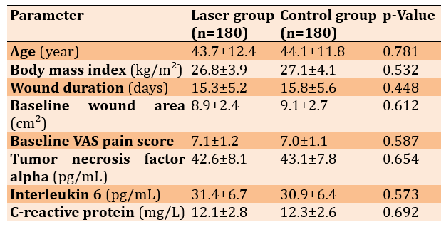
After four weeks of treatment with LLLT, systemic inflammatory biomarker levels decreased significantly. Compared with the control group, the LLLT group had lower serum concentrations of TNF-α (23.8±5.9 vs. 36.5±6.8pg/mL), IL-6 (17.2 ± 4.6 vs. 26.4±5.3pg/mL), and CRP (4.8±1.6 vs. 8.7±2.1mg/L; all p<0.001). At baseline, the mean concentrations were 42.6±8.1pg/mL for TNF-α, 31.4±6.7pg/mL for IL-6, and 12.1±2.8 mg/L for CRP. The greatest reduction was observed in CRP levels, which decreased by approximately 60% following laser treatment.
Pain intensity, tissue perfusion, complete wound-healing rates, and patient satisfaction improved significantly after PBM treatment. Compared with patients receiving standard wound care alone, those treated with LLLT had lower mean pain scores (2.4±0.9 vs. 4.5±1.2), greater tissue perfusion (34.7%± 5.1% vs. 12.8%±3.7%), and higher complete wound-healing rates (88.3% vs. 64.4%). Patient satisfaction was also higher in the LLLT group, with mean scores of 9.1±0.6 compared with 7.2±0.9 in the control group. All differences between groups were statistically significant (p<0.001).
Correlation analysis demonstrated a significant inverse association between inflammatory activity and wound healing. Serum CRP concentration showed the strongest negative correlation with the percentage of wound closure (r=−0.71, p<0.001). TNF-α and IL-6 also showed significant negative correlations with wound closure (r=−0.67 and r=−0.63, respectively; both p<0.001).
Discussion
This study aimed to investigate the efficacy of LLLT for treating skin wounds based on clinical signs, inflammatory marker levels, tissue perfusion, and pain intensity. LLLT improved wound healing, decreased inflammatory marker concentrations, increased local tissue blood flow, reduced pain, and enhanced patient satisfaction when used in combination with standard wound treatment. Thus, the aforementioned findings provide additional support for future studies demonstrating that PBM promotes tissue healing by influencing inflammatory processes and stimulating cellular regeneration.
Patients receiving LLLT demonstrated a considerably higher wound-closure rate. The experimental group experienced epithelial restoration and tissue repair more rapidly than the control group throughout the study, indicating that laser treatment is effective in enhancing the biological processes involved in wound healing. These findings are consistent with previous studies showing that PBM stimulates fibroblast proliferation, collagen production, angiogenesis, and connective-tissue remodeling, all of which contribute to faster wound closure [6-9, 17, 18]. De Freitas & Hamblin [7] stated that laser photons are absorbed by mitochondrial cytochrome c oxidase, leading to ATP production and the initiation of signals involved in cell proliferation and migration. Dompe et al. [9] summarized that PBM facilitates regenerative processes by improving cellular metabolism and vascularization.
There were significant decreases in serum TNF-α, IL-6, and CRP levels after laser treatment, indicating the positive clinical effects coincided with a reduction in systemic inflammation. Persistent elevations in pro-inflammatory markers hinder the transition from the inflammatory phase to the proliferative stage of wound healing [2, 3]. The observations regarding the anti-inflammatory properties of PBM are consistent with those of other research [8, 19, 20], showing that PBM inhibits the production of inflammatory cytokines while enhancing tissue restoration through the modulation of oxidative stress and immune-cell behavior. In addition, Gavish & Houreld [6] concluded that PBM creates a favorable microenvironment for wound healing by reducing inflammation while promoting angiogenesis.
The decrease in inflammatory markers is consistent with research conducted by our team [13].
According to Hadi et al. [14], inhibition of inflammatory processes effectively reduces the extent of tissue injury during myocardial ischemia–reperfusion. Although they emphasized the contribution of activation of the NF-κB signaling pathway to inflammatory tissue damage and oxidative stress, the decreases in TNF-α and IL-6 levels observed here indicate that suppression of inflammatory processes may also be characteristic of cutaneous wound healing.
Although these earlier findings were based on cardiovascular tissue rather than cutaneous wounds, the inflammatory mechanisms involved are likely similar in both cases. There was an increase in tissue perfusion and a substantial reduction in pain perception. Increased microcirculation resulting from laser therapy facilitates the delivery of oxygen and nutrients and the removal of waste products, thereby creating favorable conditions for tissue repair. Improved perfusion also helps fibroblasts function more efficiently and deposit collagen. Comparable results have been obtained in previous studies investigating the effects of PBM on local blood flow through vasodilation resulting from nitric oxide release and mitochondrial activation [7-9, 21].
There were significant negative relationships between inflammatory markers and wound-healing rates, with CRP showing the strongest association. Thus, patients with lower levels of inflammation may heal faster and regenerate tissue more rapidly. These results align with other findings [22, 23]. CRP is a reliable marker of the level and severity of inflammation [15, 24, 25]. Although the authors focus on inflammatory complications during pregnancy, they demonstrate the validity of CRP as a marker of systemic inflammation, confirming its potential for monitoring patients during wound healing.
Patients reported significantly higher satisfaction after undergoing PBM therapy. This observation provides clinically significant findings because patient feedback is becoming an increasingly important tool for assessing treatment outcomes. Our findings also resemble those of Yousif et al. [16], who found that patients receiving stem cell therapy experience better healing outcomes. Although the two methods differ in their biological mechanisms, they have a similar goal of improving tissue regeneration.
Some strengths of the present study include its prospective, randomized controlled design; relatively large sample size; assessment of laboratory and clinical outcomes; and incorporation of objective inflammatory markers into a noninvasive biophysical assessment. The inclusion of wound healing, inflammatory markers, tissue perfusion, and patient feedback regarding their condition provides an overall assessment of treatment effectiveness and adds clinical significance to the results.
However, several limitations need to be addressed. First, this study was conducted at a single clinic, which limits the generalizability of the findings. Second, only one wavelength and irradiation protocol were evaluated; therefore, the optimal treatment parameters could not be determined. Third, the follow-up period was limited to four weeks, which prevented assessment of long-term scarring and recurrence. Finally, other biomarkers, such as vascular endothelial growth factor (VEGF), transforming growth factor beta (TGF-β), matrix metalloproteinases (MMPs), and markers of oxidative stress, should also have been evaluated.
In summary, the results support the concept that LLLT is both safe and effective as an adjunctive intervention. By promoting faster wound closure, reducing inflammatory responses, enhancing tissue blood flow, and improving patient satisfaction, PBM represents a potentially effective approach to improving wound management. Therefore, multicenter randomized studies with extended follow-up periods and molecular assessments are needed to develop standardized treatment protocols. In general, LLLT can be considered a safe and effective method for treating superficial skin wounds and may serve as an adjunct to traditional treatment methods. Further multicenter, randomized controlled trials involving a larger number of patients, longer observation periods, and molecular assessments should be conducted to optimize laser treatment protocols and better understand the mechanisms of photobiomodulation.
Conclusion
Low-level laser therapy enhances wound healing, reduces inflammation and pain, improves tissue perfusion, and promotes better quality of life compared with traditional wound care.
Acknowledgments: The authors sincerely thank the administrative, medical, and nursing staff at the specialized dermatology and outpatient wound care clinics for their diligent assistance, patient coordination, and technical support throughout the study.
Ethical Permissions: The study protocol was reviewed and formally approved by the Institutional Research Ethics Committee of the Directorate of Education/Health Research Council (Approval Code: REC-2025-0819).
Conflicts of Interests: The authors declared no conflicts of interest.
Authors' Contribution: Yousif BG (First Author), Methodologist/Main Researcher/Discussion Writer (45%); Jabbar AA (Second Author), Introduction Writer/Assistant Researcher/Statistical Analyst (30%); Ketab AF (Third Author), Introduction Writer/Assistant Researcher (25%)
Funding/Support: This research received no specific grant or financial support from any public, commercial, or nonprofit funding agency.
Healing of skin wounds is a biological process that occurs in multiple stages, including hemostasis, inflammation, tissue regeneration, and tissue remodeling [1, 2]. The regulation of cell migration, fibroblast proliferation, neovascularization, extracellular matrix deposition, collagen synthesis, and re-epithelialization is essential to ensure efficient healing. Disruptions in the normal progression of these processes may lead to impaired healing, chronic wounds, larger scar formation, and an increased risk of infection [1, 2].
Inflammation plays a crucial role during the early phase of wound healing by eliminating pathogens and initiating tissue repair. However, excessive or prolonged inflammation delays the transition to the proliferative phase and can result in chronic wounds [2, 3]. Increased levels of pro-inflammatory cytokines, such as tumor necrosis factor alpha (TNF-α), interleukin 6 (IL-6), and C-reactive protein (CRP), as well as oxidative stress and activation of the nuclear factor kappa B (NF-κB) signaling pathway, have been associated with impaired tissue regeneration [3-5]. Therefore, the search for methods that can control inflammation and enhance tissue regeneration has become an important area of regenerative medicine.
Low-level laser therapy (LLLT), or photobiomodulation (PBM), is a noninvasive therapeutic technique that can stimulate tissue healing without causing thermal injury [6-8]. In contrast to high-power surgical lasers, LLLT uses low-energy monochromatic light that is absorbed primarily by cytochrome c oxidase in the mitochondria. This interaction enhances ATP production, regulates reactive oxygen species (ROS) release, activates fibroblast proliferation, contributes to collagen formation, promotes angiogenesis, and supports the restoration of epithelial tissue [7-9].
In addition to enhancing tissue repair, PBM has a strong anti-inflammatory effect by decreasing the levels of pro-inflammatory cytokines, improving microcirculation in the affected area, increasing tissue oxygenation, and modulating macrophage activity and extracellular matrix remodeling [8-10]. All of these biological effects create a favorable environment for wound healing and have been demonstrated in both laboratory and clinical studies [7-10].
An objective assessment of wound healing requires more than visual evaluation alone. Current wound-healing research incorporates clinical observations, inflammatory biomarkers, and noninvasive measurements. Serum levels of TNF-α, IL-6, and CRP provide quantifiable evidence of systemic inflammation, whereas assessments of tissue perfusion and temperature provide valuable information about local recovery [2, 8, 11, 12]. The use of various measurement technologies allows wound healing to be assessed more accurately than through conventional observation alone.
Studies conducted by our research group have demonstrated that targeting inflammatory pathways is biologically justified during tissue regeneration. Hadi et al. show that inhibiting apoptosis and the inflammatory response reduces tissue damage caused by myocardial ischemia-reperfusion, underscoring the need to control inflammation for successful tissue repair [13]. Furthermore, the findings of Hadi et al. shed light on the role of NF-κB-mediated oxidative pathways, confirming that manipulating these pathways can improve regenerative outcomes [14]. Sadiq et al. further support the findings of Hadi et al. by emphasizing the clinical relevance of CRP as a biomarker of inflammation [15]. In addition, Yousif et al. reported that regenerative treatments improve outcomes in patients receiving stem cell transplants [16].
Although many studies have been conducted on PBM, significant inconsistencies remain regarding laser wavelength, energy density, irradiation method, treatment duration, and outcome assessment [6-10]. Consequently, clinical outcomes are often contradictory, and no standardized therapeutic protocols currently exist. Furthermore, only a few clinical trials have analyzed the effects of the aforementioned procedures on wound healing together with patients’ inflammatory biomarkers and physical parameters.
This study aimed to evaluate the efficacy of LLLT in addition to standard wound care by using clinical evaluations, laboratory analyses of inflammatory biomarkers, and noninvasive assessments of physical parameters. By simultaneously examining wound closure, inflammatory processes, tissue perfusion, pain, and patient-centered outcomes, this study is expected to clarify the benefits of PBM for wound healing and tissue regeneration.
Materials and Methods
Design and sample
This controlled, prospective, randomized clinical study was conducted at the outpatient wound treatment and dermatology clinics of a specialized medical center over a six-month period. The study included clinical, laboratory, and noninvasive biophysical evaluations. Initially, 360 individuals with superficial or partial-thickness wounds were recruited and randomly assigned in a 1:1 ratio to two groups. Group A, the intervention group, included 180 individuals who received low-level laser therapy (LLLT) in addition to standard wound care, whereas Group B, the control group, included 180 individuals who received standard wound care alone.
Sample size was calculated using G*Power 3.1.9.7. Based on previous clinical trials of low-level laser therapy for cutaneous wound healing, an anticipated effect size (Cohen’s d) of 0.35 was assumed for the primary outcome: the percentage of wound closure 4 weeks after the intervention. With an alpha level (α) of 0.05 and 90% statistical power (1−β=0.9) for a two-tailed independent-samples t test, the minimum required sample size was estimated at approximately 172 participants per arm. Allowing for potential dropout, attrition, and noncompliance during the 4-week protocol (approximately 5%), 360 participants (180 per group) were recruited.
The inclusion criteria were age between 18 and 65 years; presence of a superficial or partial-thickness wound with a duration of 7–30 days; wound size suitable for digital measurement; ability to attend the treatment sessions; provision of written informed consent; absence of uncontrolled diabetes mellitus, severe systemic infection, malignant skin tumors, autoimmune skin conditions, photosensitivity disorders, pregnancy or breastfeeding, previous laser treatment at the lesion site, current use of immunosuppressants, and serious injuries requiring surgical treatment.
Procedure
Written informed consent was obtained from all participants before enrollment. Participant confidentiality was maintained, and the research procedures followed the ethical principles of the Declaration of Helsinki and Good Clinical Practice.
In addition to standard wound care, participants in the experimental group received LLLT using a gallium-aluminum-arsenide (GaAlAs) continuous-wave diode laser. The laser operated at a wavelength of 660 nm and an output power of 100mW (0.1W). The circular aperture beam had a spot size of 0.5cm², corresponding to an irradiance of 200mW/cm² (0.2 W/cm²). An energy density (fluence) of 4.0J/cm² was delivered to each irradiated point. Accordingly, the exposure time was 20 seconds per point (4.0J/cm² ÷ 0.2W/cm²=20 seconds).
The laser was applied three times per week for four weeks using a perpendicular, noncontact technique. The applicator was held at a consistent distance of 0.5 cm from the wound surface under strict sterile conditions. A structured multipoint grid technique was used, with irradiation points distributed across the wound bed at 1-cm intervals. Additional circumferential points were placed at 1-cm intervals along the intact periwound margins to promote vascularization of the granulation-tissue bed and peripheral epithelial cell migration. The dose was adjusted according to the baseline wound area. For an average wound area of approximately 9cm², approximately 18 nonoverlapping points were treated, resulting in a total delivered optical energy of approximately 36J per session (2J per point). Both the patient and the staff member performing the procedure wore laser-protection glasses during each application.
All participants received the same standardized wound care. The wound bed was cleansed with sterile normal saline, and necrotic tissue was removed when necessary. A sterile wound dressing was then applied. Topical agents, including growth factors, biological dressings, platelet-rich plasma, and advanced wound-healing drugs, were not used to minimize bias.
Clinical evaluations were performed before treatment began and once weekly during the four-week treatment period. Standardized photographs were taken using the same camera settings, lighting conditions, and distance from the wound. Digital wound-measurement techniques were used to determine the wound area. Clinical outcomes included the percentage of wound closure, local erythema, edema, wound exudate, pain in the affected area, and complete wound healing.
An objective biophysical evaluation was conducted under controlled environmental conditions. Skin temperature surrounding the wound was measured using thermal imaging or an infrared thermometer. Tissue perfusion was assessed using laser Doppler flowmetry, when available. Measurements were obtained before therapy began and after therapy was completed.
Venous blood samples were collected at baseline and after four weeks of therapy. Serum was separated and analyzed according to standard laboratory procedures. Inflammatory biomarkers, including TNF-α, IL-6, and CRP, were quantified. Serum TNF-α and IL-6 levels were determined using enzyme-linked immunosorbent assay (ELISA) kits according to the manufacturers’ instructions, whereas CRP levels were measured using routine automated clinical chemistry techniques.
Pain severity was measured using the 10-point visual analog scale (VAS), with 0 indicating no pain and 10 indicating the most severe pain imaginable. Patient satisfaction with the treatment outcome was assessed at the end of the treatment period using a satisfaction questionnaire, with higher scores indicating greater satisfaction.
Statistical analysis
SPSS 26 was used for statistical analysis. The t-test was used to compare parameters between and within the two groups. The chi-square test was used to compare categorical parameters. The association between the percentage of wound closure and inflammatory markers was determined using Pearson’s correlation coefficients.
Findings
A total of 360 patients were included in the study. No patients were lost during the follow-up period, and all completed the treatment protocol. In the laser group, 98 participants (54.4%) were male and 82 (45.6%) were female. In the control group, 95 participants (52.8%) were male and 85 (47.2%) were female. The distribution of sex did not differ significantly between the groups (p=0.764). No statistically significant differences were observed between the study groups at baseline (p>0.05; Table 1).
Table 1. Mean baseline demographic and clinical characteristics of the participants
After four weeks of treatment with LLLT, systemic inflammatory biomarker levels decreased significantly. Compared with the control group, the LLLT group had lower serum concentrations of TNF-α (23.8±5.9 vs. 36.5±6.8pg/mL), IL-6 (17.2 ± 4.6 vs. 26.4±5.3pg/mL), and CRP (4.8±1.6 vs. 8.7±2.1mg/L; all p<0.001). At baseline, the mean concentrations were 42.6±8.1pg/mL for TNF-α, 31.4±6.7pg/mL for IL-6, and 12.1±2.8 mg/L for CRP. The greatest reduction was observed in CRP levels, which decreased by approximately 60% following laser treatment.
Pain intensity, tissue perfusion, complete wound-healing rates, and patient satisfaction improved significantly after PBM treatment. Compared with patients receiving standard wound care alone, those treated with LLLT had lower mean pain scores (2.4±0.9 vs. 4.5±1.2), greater tissue perfusion (34.7%± 5.1% vs. 12.8%±3.7%), and higher complete wound-healing rates (88.3% vs. 64.4%). Patient satisfaction was also higher in the LLLT group, with mean scores of 9.1±0.6 compared with 7.2±0.9 in the control group. All differences between groups were statistically significant (p<0.001).
Correlation analysis demonstrated a significant inverse association between inflammatory activity and wound healing. Serum CRP concentration showed the strongest negative correlation with the percentage of wound closure (r=−0.71, p<0.001). TNF-α and IL-6 also showed significant negative correlations with wound closure (r=−0.67 and r=−0.63, respectively; both p<0.001).
Discussion
This study aimed to investigate the efficacy of LLLT for treating skin wounds based on clinical signs, inflammatory marker levels, tissue perfusion, and pain intensity. LLLT improved wound healing, decreased inflammatory marker concentrations, increased local tissue blood flow, reduced pain, and enhanced patient satisfaction when used in combination with standard wound treatment. Thus, the aforementioned findings provide additional support for future studies demonstrating that PBM promotes tissue healing by influencing inflammatory processes and stimulating cellular regeneration.
Patients receiving LLLT demonstrated a considerably higher wound-closure rate. The experimental group experienced epithelial restoration and tissue repair more rapidly than the control group throughout the study, indicating that laser treatment is effective in enhancing the biological processes involved in wound healing. These findings are consistent with previous studies showing that PBM stimulates fibroblast proliferation, collagen production, angiogenesis, and connective-tissue remodeling, all of which contribute to faster wound closure [6-9, 17, 18]. De Freitas & Hamblin [7] stated that laser photons are absorbed by mitochondrial cytochrome c oxidase, leading to ATP production and the initiation of signals involved in cell proliferation and migration. Dompe et al. [9] summarized that PBM facilitates regenerative processes by improving cellular metabolism and vascularization.
There were significant decreases in serum TNF-α, IL-6, and CRP levels after laser treatment, indicating the positive clinical effects coincided with a reduction in systemic inflammation. Persistent elevations in pro-inflammatory markers hinder the transition from the inflammatory phase to the proliferative stage of wound healing [2, 3]. The observations regarding the anti-inflammatory properties of PBM are consistent with those of other research [8, 19, 20], showing that PBM inhibits the production of inflammatory cytokines while enhancing tissue restoration through the modulation of oxidative stress and immune-cell behavior. In addition, Gavish & Houreld [6] concluded that PBM creates a favorable microenvironment for wound healing by reducing inflammation while promoting angiogenesis.
The decrease in inflammatory markers is consistent with research conducted by our team [13].
According to Hadi et al. [14], inhibition of inflammatory processes effectively reduces the extent of tissue injury during myocardial ischemia–reperfusion. Although they emphasized the contribution of activation of the NF-κB signaling pathway to inflammatory tissue damage and oxidative stress, the decreases in TNF-α and IL-6 levels observed here indicate that suppression of inflammatory processes may also be characteristic of cutaneous wound healing.
Although these earlier findings were based on cardiovascular tissue rather than cutaneous wounds, the inflammatory mechanisms involved are likely similar in both cases. There was an increase in tissue perfusion and a substantial reduction in pain perception. Increased microcirculation resulting from laser therapy facilitates the delivery of oxygen and nutrients and the removal of waste products, thereby creating favorable conditions for tissue repair. Improved perfusion also helps fibroblasts function more efficiently and deposit collagen. Comparable results have been obtained in previous studies investigating the effects of PBM on local blood flow through vasodilation resulting from nitric oxide release and mitochondrial activation [7-9, 21].
There were significant negative relationships between inflammatory markers and wound-healing rates, with CRP showing the strongest association. Thus, patients with lower levels of inflammation may heal faster and regenerate tissue more rapidly. These results align with other findings [22, 23]. CRP is a reliable marker of the level and severity of inflammation [15, 24, 25]. Although the authors focus on inflammatory complications during pregnancy, they demonstrate the validity of CRP as a marker of systemic inflammation, confirming its potential for monitoring patients during wound healing.
Patients reported significantly higher satisfaction after undergoing PBM therapy. This observation provides clinically significant findings because patient feedback is becoming an increasingly important tool for assessing treatment outcomes. Our findings also resemble those of Yousif et al. [16], who found that patients receiving stem cell therapy experience better healing outcomes. Although the two methods differ in their biological mechanisms, they have a similar goal of improving tissue regeneration.
Some strengths of the present study include its prospective, randomized controlled design; relatively large sample size; assessment of laboratory and clinical outcomes; and incorporation of objective inflammatory markers into a noninvasive biophysical assessment. The inclusion of wound healing, inflammatory markers, tissue perfusion, and patient feedback regarding their condition provides an overall assessment of treatment effectiveness and adds clinical significance to the results.
However, several limitations need to be addressed. First, this study was conducted at a single clinic, which limits the generalizability of the findings. Second, only one wavelength and irradiation protocol were evaluated; therefore, the optimal treatment parameters could not be determined. Third, the follow-up period was limited to four weeks, which prevented assessment of long-term scarring and recurrence. Finally, other biomarkers, such as vascular endothelial growth factor (VEGF), transforming growth factor beta (TGF-β), matrix metalloproteinases (MMPs), and markers of oxidative stress, should also have been evaluated.
In summary, the results support the concept that LLLT is both safe and effective as an adjunctive intervention. By promoting faster wound closure, reducing inflammatory responses, enhancing tissue blood flow, and improving patient satisfaction, PBM represents a potentially effective approach to improving wound management. Therefore, multicenter randomized studies with extended follow-up periods and molecular assessments are needed to develop standardized treatment protocols. In general, LLLT can be considered a safe and effective method for treating superficial skin wounds and may serve as an adjunct to traditional treatment methods. Further multicenter, randomized controlled trials involving a larger number of patients, longer observation periods, and molecular assessments should be conducted to optimize laser treatment protocols and better understand the mechanisms of photobiomodulation.
Conclusion
Low-level laser therapy enhances wound healing, reduces inflammation and pain, improves tissue perfusion, and promotes better quality of life compared with traditional wound care.
Acknowledgments: The authors sincerely thank the administrative, medical, and nursing staff at the specialized dermatology and outpatient wound care clinics for their diligent assistance, patient coordination, and technical support throughout the study.
Ethical Permissions: The study protocol was reviewed and formally approved by the Institutional Research Ethics Committee of the Directorate of Education/Health Research Council (Approval Code: REC-2025-0819).
Conflicts of Interests: The authors declared no conflicts of interest.
Authors' Contribution: Yousif BG (First Author), Methodologist/Main Researcher/Discussion Writer (45%); Jabbar AA (Second Author), Introduction Writer/Assistant Researcher/Statistical Analyst (30%); Ketab AF (Third Author), Introduction Writer/Assistant Researcher (25%)
Funding/Support: This research received no specific grant or financial support from any public, commercial, or nonprofit funding agency.
Keywords:
References
1. Sen CK. Human wounds and its burden: An updated compendium of estimates. Adv Wound Care. 2019;8(2):39-48. [Link] [DOI:10.1089/wound.2019.0946]
2. Rodrigues M, Kosaric N, Bonham CA, Gurtner GC. Wound healing: A cellular perspective. Physiol Rev. 2019;99(1):665-706. [Link] [DOI:10.1152/physrev.00067.2017]
3. Eming SA, Wynn TA, Martin P. Inflammation and metabolism in tissue repair and regeneration. Science. 2017;356(6342):1026-30. [Link] [DOI:10.1126/science.aam7928]
4. Landén NX, Li D, Ståhle M. Transition from inflammation to proliferation: A critical step during wound healing. Cell Mol Life Sci. 2016;73(20):3861-85. [Link] [DOI:10.1007/s00018-016-2268-0]
5. Guo S, Dipietro LA. Factors affecting wound healing. J Dent Res. 2010;89(3):219-29. [Link] [DOI:10.1177/0022034509359125]
6. Gavish L, Houreld NN. Photobiomodulation therapy for wound healing and skin regeneration. Adv Skin Wound Care. 2019;32(8):361-4. [Link]
7. De Freitas LF, Hamblin MR. Proposed mechanisms of photobiomodulation or low-level light therapy. IEEE J Sel Top Quantum Electron. 2016;22(3):7000417. [Link] [DOI:10.1109/JSTQE.2016.2561201]
8. Hamblin MR. Mechanisms and applications of the anti-inflammatory effects of photobiomodulation. AIMS Biophys. 2017;4(3):337-61. [Link] [DOI:10.3934/biophy.2017.3.337]
9. Dompe C, Moncrieff L, Matys J, Grzech-Leśniak K, Kocherova I, Bryja A, et al. Photobiomodulation-underlying mechanisms and clinical applications. J Clin Med. 2020;9(6):1724. [Link] [DOI:10.3390/jcm9061724]
10. Aggarwal I, Lio PA. Photobiomodulation therapy and low-level light therapy in wound healing. Lasers Med Sci. 2023;38(1):239. [Link] [DOI:10.1007/s10103-023-03909-9]
11. Popko K, Gorska E, Stelmaszczyk-Emmel A, Plywaczewski R, Stoklosa A, Gorecka D, et al. Proinflammatory cytokines Il-6 and TNF-α and the development of inflammation in obese subjects. Eur J Med Res. 2010;15(Suppl 2):120. [Link] [DOI:10.1186/2047-783X-15-S2-120]
12. Florescu DN, Boldeanu MV, Șerban RE, Florescu LM, Serbanescu MS, Ionescu M, et al. Correlation of the pro-inflammatory cytokines IL-1β, IL-6, and TNF-α, inflammatory markers, and tumor markers with the diagnosis and prognosis of colorectal cancer. Life. 2023;13(12):2261. [Link] [DOI:10.3390/life13122261]
13. Hadi NR, Al-Amran FG, Yousif MG, Alrekabi MD, Hussein FH. Amelioration of myocardial ischaemia/reperfusion injury by L-carnitine via down-regulation of the inflammatory response and apoptosis. Exp Clin Cardiol. 2014;20(8):3732-40. [Link]
14. Hadi NR, Yousif NG, Abdulzahra MS, Mohammad BI, Al-Amran FG, Majeed ML, et al. Role of NF-κB and oxidative pathways in atherosclerosis: Cross-talk between dyslipidaemia and candesartan. Cardiovasc Ther. 2013;31(6):381-7. [Link] [DOI:10.1111/1755-5922.12033]
15. Sadiq AM, Hussein CM, Mohammed RJ, Yousif MG. Correlation between highly sensitive C-reactive protein level in cases of preeclampsia with or without intrauterine growth restriction. Indian J Forensic Med Toxicol. 2020;14(4):6012-6. [Link]
16. Yousif NG, Yousif MG, Mohsen AAU, El-Bakaa HS, Younise MH, Altimimi AN, et al. Prospective single center analysis of outcome stem cell transplants in patients with cerebral palsy. Pol Merkur Lekarski. 2023;51(4):339-45. [Link] [DOI:10.36740/Merkur202304107]
17. Cavalcanti MF, Cabette RA, Moraes AC, Diomede F, Trubiani O, Maria DA. The effects in vitro of photobiomodulation over fibroblasts and extracellular matrix. Photobiomodul Photomed Laser Surg. 2024;42(2):140-7. [Link] [DOI:10.1089/photob.2023.0123]
18. Jarmołowicz M, Kotela A, Laszczyńska M, Wesołek K, Gajewska M, Błaszczyk-Pośpiech A, et al. LED-based photobiomodulation in fibroblast and osteoblast models: a systematic review of in vitro evidence. Appl Sci. 2026;16(17):8399. [Link] [DOI:10.3390/app16178399]
19. Shamloo S, Defensor E, Ciari P, Ogawa G, Vidano L, Lin JS, et al. The anti-inflammatory effects of photobiomodulation are mediated by cytokines: Evidence from a mouse model of inflammation. Front Neurosci. 2023;17:1150156. [Link] [DOI:10.3389/fnins.2023.1150156]
20. Ryu JH, Park J, Kim BY, Kim Y, Kim NG, Shin YI. Photobiomodulation ameliorates inflammatory parameters in fibroblast-like synoviocytes and experimental animal models of rheumatoid arthritis. Front Immunol. 2023;14:1122581. [Link] [DOI:10.3389/fimmu.2023.1122581]
21. Lai F, Mai Y, Wang Z, Wang C, Xu S. Photobiomodulation Targets Mitochondrial Homeostasis for Diabetic Wound Healing. Tissue Eng. Part B Rev. 2026:19373368261460337. [Link] [DOI:10.1177/19373368261460337]
22. Xu F, Zhang C, Graves DT. Abnormal cell responses and role of TNF‐α in impaired diabetic wound healing. BioMed Res Int. 2013;2013(1):754802. [Link] [DOI:10.1155/2013/754802]
23. Čoma M, Fröhlichová L, Urban L, Zajíček R, Urban T, Szabo P, et al. Molecular changes underlying hypertrophic scarring following burns involve specific deregulations at all wound healing stages (inflammation, proliferation and maturation). Int J Mol Sci. 2021;22(2):897. [Link] [DOI:10.3390/ijms22020897]
24. Germolec DR, Shipkowski KA, Frawley RP, Evans E. Markers of inflammation. Immunotoxicit Test. 2018:57-79. [Link] [DOI:10.1007/978-1-4939-8549-4_5]
25. Hessam S, Sand M, Gambichler T, Bechara FG. Correlation of inflammatory serum markers with disease severity in patients with hidradenitis suppurativa (HS). J Am Acad Dermatol. 2015;73(6):998-1005. [Link] [DOI:10.1016/j.jaad.2015.08.052]