

JMERC
Owner
Janbazan Medical and Engineering Research Center (JMERC) is the owner of IJWPH.

1.0
Cite Score 2025
SJR: 0.129 / SNIP: 0.140
Volume 18, Issue 2 (2026)
Iran J War Public Health 2026, 18(2): 249-255 |
Back to browse issues page
Article Type:
Subject:




History
Received: 2026/06/15 | Accepted: 2026/08/3 | Published: 2026/08/21
Received: 2026/06/15 | Accepted: 2026/08/3 | Published: 2026/08/21
How to cite this article
Jasim A, Hammadi A, Barton S. Comparative Diagnostic Methods for Identifying Bacterial Respiratory Pathogens. Iran J War Public Health 2026; 18 (2) :249-255
URL: http://ijwph.ir/article-1-1805-en.html
URL: http://ijwph.ir/article-1-1805-en.html
Download citation:
BibTeX | RIS | EndNote | Medlars | ProCite | Reference Manager | RefWorks
Send citation to:


Rights and permissions
BibTeX | RIS | EndNote | Medlars | ProCite | Reference Manager | RefWorks
Send citation to:

1- Department of Pathological Analysis, College of Applied Medical Sciences, University of Kerbala, Karbala, Iraq
2- Department of Analytical Chemistry, Faculty of Pharmaceutics, Kingston University, London, England
2- Department of Analytical Chemistry, Faculty of Pharmaceutics, Kingston University, London, England
Full-Text (HTML) (6 Views)
Introduction
Respiratory tract infections (RTIs) are among the leading causes of morbidity and mortality worldwide and remain major reasons for hospital admission and antibiotic use [1, 2]. Lower respiratory tract infections (LRTIs) are the fourth leading cause of death globally and the most common infectious cause of mortality, accounting for approximately 2.4 million deaths annually [3]. In Iraq, the burden of RTIs is particularly high.
A pediatric study conducted in Kerbala reported that LRTIs account for 71.7% of respiratory infections among hospitalized children, with more than one-quarter of cases requiring admission to an intensive care unit [4]. During the COVID-19 pandemic, secondary bacterial infections further increased mortality rates among hospitalized patients in Iraq, emphasizing the need for rapid and reliable diagnostic methods [5]. Despite the significant burden of infectious diseases, diagnostic resources in many developing countries, including Iraq, remain limited because of inadequate healthcare funding. Consequently, clinicians often administer empirical antibiotic therapy before confirming the causative pathogen. This practice has contributed to the increasing emergence of multidrug-resistant organisms (MDROs), including methicillin-resistant Staphylococcus aureus (MRSA) and carbapenem-resistant Klebsiella pneumoniae [6, 7].
Accurate and rapid diagnostic techniques could improve patient management by enabling early targeted therapy, reducing the unnecessary use of broad-spectrum antibiotics, and decreasing healthcare costs and the duration of hospital stays [8]. Bacterial pathogens, such as Streptococcus pneumoniae, K. pneumoniae, S. aureus, and Haemophilus influenzae are among the most frequently isolated organisms in both community-acquired and hospital-acquired respiratory infections [9]. Traditionally, sputum culture has been considered the gold standard for bacterial diagnosis. Conventional culture methods, combined with biochemical testing or automated systems such as VITEK® 2, can identify bacterial species and determine antimicrobial susceptibility profiles [10]. However, these methods have several important limitations. Culture requires viable organisms, and prior antibiotic administration may lead to false-negative results. In addition, fastidious organisms, such as S. pneumoniae and H. influenzae require special growth conditions, while definitive culture results may take 48–72 hours, delaying appropriate treatment decisions [11].
Molecular diagnostic techniques, particularly polymerase chain reaction (PCR) and real-time PCR, offer several advantages over conventional methods. PCR can directly detect bacterial DNA in clinical specimens without requiring viable organisms and can provide rapid, highly sensitive, and specific results. Multiplex PCR assays can simultaneously detect multiple respiratory pathogens within a short period [12]. PCR can detect significantly more cases of pathogens, such as H. influenzae and S. pneumoniae, than culture, especially in patients who received antibiotics before sampling [13]. Furthermore, PCR can identify difficult-to-culture organisms, including Mycoplasma pneumoniae and Mycobacterium tuberculosis, and may detect antimicrobial resistance genes that can guide antibiotic selection [14]. Nevertheless, molecular techniques cannot completely replace conventional culture methods. PCR may detect DNA from nonviable organisms, potentially resulting in false-positive findings, and it does not provide comprehensive phenotypic antimicrobial susceptibility testing, which still depends on cultured isolates [15]. In addition, the high cost and technical requirements of molecular assays may limit their routine use in resource-limited healthcare settings [16-19].
This study aimed to compare conventional culture methods, automated identification using VITEK® 2, and species-specific PCR to detect bacterial pathogens in respiratory samples obtained from 180 clinically ill Iraqi patients. We aimed to evaluate the prevalence and distribution of bacterial pathogens identified by each method, assess the agreement between conventional and molecular techniques, and determine the additional diagnostic value of PCR, particularly for S. pneumoniae and M. tuberculosis. The findings may contribute to the development of evidence-based diagnostic strategies for RTIs in Iraq.
Materials and Methods
Design and sampling
This cross-sectional observational study was conducted at Samarra General Hospital and Baghdad Medical City in Iraq from October 2025 to February 2026. We recruited 180 patients (110 males and 70 females) aged 2-50 years who had clinical signs of RTIs, such as cough, fever, sputum production, and dyspnea.
Patients were included if they had symptomatic respiratory infections, available clinical and/or demographic data, and respiratory specimens collected from the appropriate site at the appropriate time while they were experiencing respiratory symptoms.
Sputum specimens and nasopharyngeal swabs were obtained from all participants using sterile collection materials and separate bacterial and viral transport systems to prevent cross-contamination. Sputum specimens were collected directly from the patients and immediately transported to the laboratory within 1–3 hours of collection. All samples were stored at 2–4°C to maintain bacterial viability, and aliquots designated for molecular analysis were stored at −20°C to ensure nucleic acid integrity. The samples were properly labeled with patient identification and collection time to facilitate sample tracking and analysis.
Microscopic diagnosis of Pulmonary Tuberculosis (Ziehl-Neelsen Stain)
Microscopic examination of respiratory samples for M. tuberculosis was performed using the Ziehl–Neelsen (acid-fast) staining method. Other bacteria were examined microscopically using Gram staining.
Bacterial culture
Blood agar and MacConkey agar were used for bacterial isolation and identification. Blood agar served as an enriched medium for the growth of fastidious bacteria and the detection of hemolytic activity, particularly among Gram-positive organisms. MacConkey agar was used as a selective and differential medium for the isolation of Gram-negative bacteria. Lowenstein–Jensen (LJ) medium is a selective and enriched solid medium specifically used for the isolation and cultivation of M. tuberculosis.
For Media preparation, blood agar and MacConkey agar were prepared according to the manufacturer’s instructions (HiMedia Laboratories Pvt. Ltd., Mumbai, India) and Lowenstein–Jensen medium was prepared according to the manufacturer’s instructions (HiMedia Laboratories Pvt. Ltd., Mumbai, India).
The VITEK® 2 Compact System (bioMérieux, France) was used for bacterial identification.
Molecular detection (PCR)
The ABIOpure™ Total DNA Extraction Kit (ABIOpure, USA) was used for bacterial DNA extraction according to the manufacturer’s instructions. The target organisms were S. pneumoniae, S. aureus, K. pneumoniae, and M. tuberculosis (Table 1). Reactions were assembled in a total volume of 50µL containing GoTaq Green Master Mix (Promega, USA), forward and reverse primers (10µM), template DNA, and nuclease-free water. Thermal cycling consisted of an initial denaturation at 95°C for 5 minutes, followed by 30 cycles of denaturation at 95°C for 30 seconds, annealing at 54°C for 30 seconds, and extension at 72°C for 30 seconds, followed by a final extension at 72°C for 7 minutes.
The study was conducted in accordance with the ethical principles for research involving human participants. Written informed consent was obtained from the participants or their legal guardians, where applicable.
Table 1. Sequences of primers for bacterial detection

Statistical analysis
Data were statistically analyzed using SPSS 23. The chi-square test or Fisher’s exact test was used to determine associations between categorical parameters, such as smoking status, infection status, age, and sex.
Findings
A total of 180 clinical respiratory samples were collected from patients. Of these, 150 (83.3%) were confirmed to be infected using microbiological and molecular diagnostic methods, whereas 30 (16.7%) were noninfected despite clinical suspicion of infection. Among females, 55 of 70 samples (78.6%) were positive, compared with 95 of 110 samples (86.4%) among males; however, this difference was not statistically significant (p=0.13). By age group, infection rates were 75.0% among patients younger than 5 years, 80.0% among those aged 5–15 years, 83.3% among those aged 16–25 years, 84.4% among those aged 30–35 years, and 85.7% among those older than 50 years. No statistically significant association was observed between age and infection status (overall p≈0.84).
A statistically significant association was observed between smoking status and respiratory tract infection (p<0.0001). Of the 60 smokers, 40 (66.7%) had a respiratory tract infection, compared with 110 (91.7%) of the 120 nonsmokers. Among all infected participants, smokers represented 26.7% (40/150), whereas nonsmokers represented 73.3% (110/150). Chronic diseases were also significantly associated with respiratory tract infection according to the reported analysis (p<0.0001). Hypertension was present in 30 participants, corresponding to 16.7% of the total study population, while diabetes mellitus was present in 10 participants, corresponding to 5.6% of the total population (p<0.0001). Overall, smoking status, hypertension, and diabetes mellitus showed statistically significant relationships with RTIs.
Culture using blood agar and MacConkey agar most frequently identified K. pneumoniae (26.7%), S. pneumoniae (20.0%), and S. aureus (18.7%), while additional less common organisms were also recovered. M. tuberculosis was not detected on these routine media because they are unsuitable for mycobacterial growth; however, LJ culture identified it in 20 cases (13.3%). PCR detected the selected target pathogens, with S. pneumoniae being the most frequent (41.3%), followed by S. aureus (28.0%), K. pneumoniae (17.3%), and M. tuberculosis (13.3%). PCR detected significantly more S. pneumoniae than VITEK® 2 (41.3% vs. 20.0%; p < 0.001). VITEK® 2 identified significantly more K. pneumoniae than PCR (26.7% vs. 17.3%; p=0.004). M. tuberculosis was detected by PCR but not by VITEK® 2 (p<0.001). Overall, VITEK® 2 identified the cultured bacterial isolates and produced findings similar to routine culture, but it did not identify M. tuberculosis (Table 2).
S. aureus produced dense, opaque, cream-colored
colonies with clear zones of β-hemolysis on blood agar, whereas S. pneumoniae formed small gray colonies with surrounding greenish α-hemolysis. On MacConkey agar, K. pneumoniae appeared as large, mucoid, pink colonies, consistent with lactose fermentation. M. tuberculosis was detected only on Lowenstein–Jensen medium, where its growth confirms the recovery of 20 culture-positive cases; it does not grow on routine blood or MacConkey agar (Figure 1).
Table 2. Frequency of bacterial pathogens detected by culture, polymerase chain reaction, and VITEK® 2
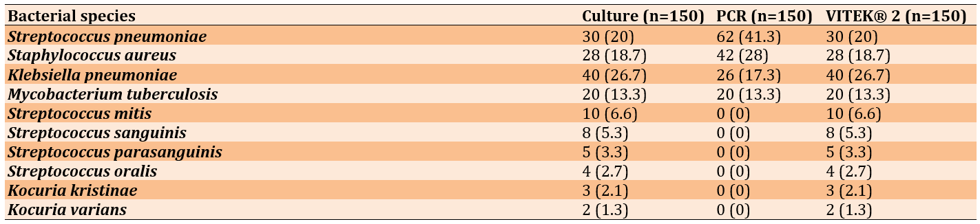
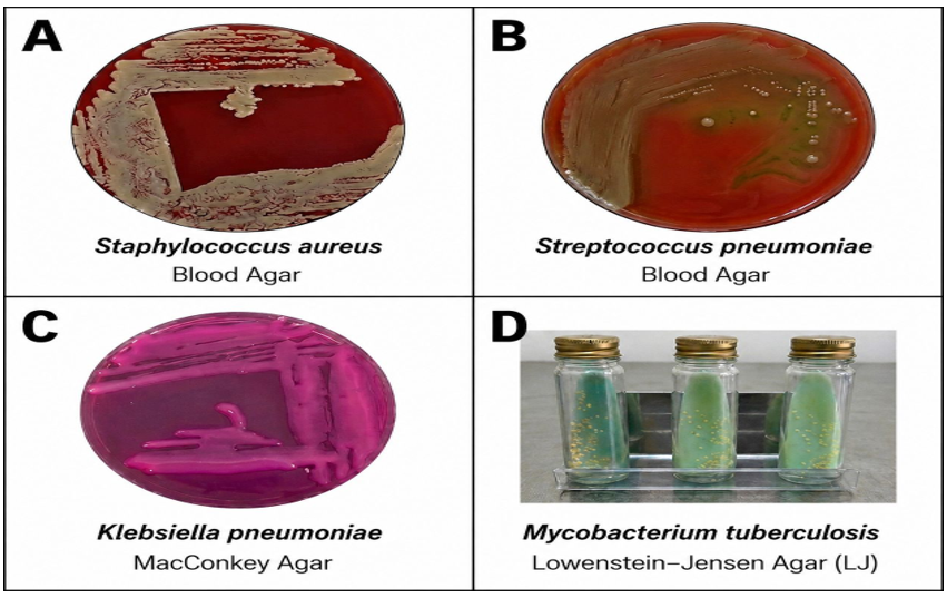
Figure 1. Growth characteristics of bacterial isolates on different culture media. (A) Staphylococcus aureus on blood agar; (B) Streptococcus pneumoniae on blood agar; (C) Klebsiella pneumoniae on MacConkey agar; and (D) Mycobacterium tuberculosis on Lowenstein–Jensen (LJ) medium.
The distinct DNA bands observed at the expected sizes—100 bp for S. aureus, 87bp for M. tuberculosis, 230bp for S. pneumoniae, and 144bp for K. pneumoniae—indicate successful amplification of the corresponding targets. The absence of bands in the non-target strains supports the specificity of the PCR assays. Together, PCR provided a rapid method for confirming pathogen identity and detecting M. tuberculosis, which requires specialized culture media (Figure 2).
Single-, dual-, and triple-pathogen infections were identified in 103 (57.2%), 45 (25.0%), and 25 (13.9%) cases, respectively. No pathogen was detected in 7 cases (3.9%).
Bacterial-only infections occurred in 120 cases (66.7%), mixed bacterial–viral infections in 33 (18.3%), and viral-only infections in 17 (9.4%). Overall, the distributions were statistically significant (p<0.001), except for dual-pathogen infections (p=1.00).
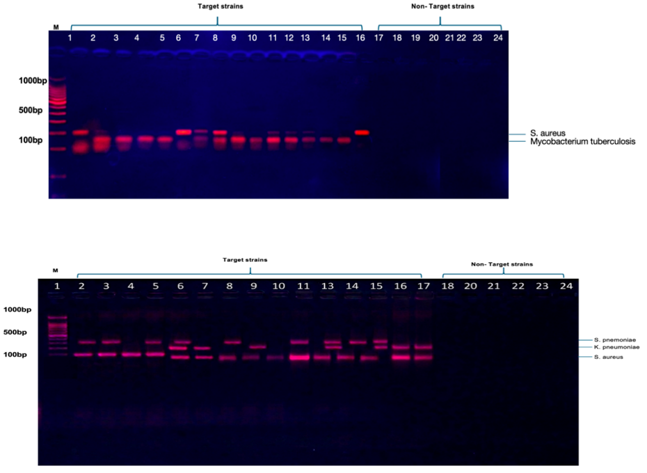
Figure 2. Agarose gel electrophoresis of PCR products from bacterial pathogens. (A) Detection of Staphylococcus aureus (100 bp) and Mycobacterium tuberculosis (87 bp). Lane M, 100-bp DNA ladder; lane 2, Staphylococcus aureus; lane 3, Mycobacterium tuberculosis; lanes 4–24, non-target strains (negative controls). (B) Detection of Streptococcus pneumoniae (230 bp), Klebsiella pneumoniae (144 bp), and Staphylococcus aureus (100 bp). Lanes 1–3 correspond to Streptococcus pneumoniae, Klebsiella pneumoniae, and Staphylococcus aureus, respectively; lane M, 100-bp DNA ladder; lanes 4–24, non-target strains (negative controls). Distinct bands at the expected amplicon sizes indicate successful amplification.
Discussion
This study for the first in Iraq compared conventional culture, VITEK® 2 automated identification, and species-specific PCR for diagnosing bacterial respiratory pathogens within the same clinical cohort. There was a high prevalence of bacterial respiratory infections among symptomatic patients (83.3%) and each diagnostic method had distinct advantages and limitations. PCR proved superior for detecting S. pneumoniae and M. tuberculosis, whereas culture-based methods remained important for identifying opportunistic and commensal organisms and for performing antimicrobial susceptibility testing. The overall infection rate observed is consistent with previous Iraqi studies reporting a high burden of bacterial respiratory infections, particularly among hospitalized and critically ill patients [20-23]. The predominance of pathogens, such as K. pneumoniae, S. pneumoniae, and P. aeruginosa supports regional surveillance data identifying these bacteria as major causes of RTIs and hospital-acquired pneumonia in the Middle East [24, 25].
A significantly higher detection rate of S. pneumoniae by PCR compared with the culture and VITEK® 2 methods was observed. PCR identified the organism in 41.3% of infected samples, whereas VITEK® 2 detected it in only 20.0%. This ovservation is consistent with previous studies showing that molecular techniques are more sensitive, particularly in patients who received antibiotics before sample collection [26]. The low recovery of pneumococci by culture may be attributed to organism autolysis, fastidious growth requirements, and suppression by prior antibiotic therapy [27]. These findings support incorporating PCR into routine diagnostic protocols for S. pneumoniae, especially in cases with negative cultures despite strong clinical suspicion.
PCR also demonstrated substantial diagnostic value for M. tuberculosis. Although no isolates grew on routine blood agar or MacConkey agar, 20 cases (13.3%) were successfully detected on Lowenstein-Jensen (LJ) medium and confirmed by Ziehl–Neelsen staining and PCR targeting the EsxA gene. All LJ culture-positive samples were PCR-positive, demonstrating complete agreement between the two methods [28-30]. However, specialized mycobacterial culture facilities are unavailable in many Iraqi healthcare settings, and diagnosis often relies solely on smear microscopy and clinical assessment. Therefore, routine implementation of PCR could significantly improve tuberculosis detection and reduce disease transmission [31, 32].
In contrast, culture and VITEK® 2 identified K. pneumoniae more frequently than PCR. This discrepancy may be related to primer variability, a low bacterial load, or upper respiratory tract colonization rather than active infection. Nevertheless, the high prevalence of K. pneumoniae is consistent with reports describing it as a leading multidrug-resistant respiratory pathogen in Iraq and neighboring countries [33, 34]. Consequently, conventional culture remains essential for detecting this organism and performing antimicrobial susceptibility testing.
Another important finding was that culture-based methods can detect opportunistic and commensal organisms not included among the PCR targets, including Streptococcus mitis, S. oralis, and Kocuria species. Although these organisms are part of the normal oropharyngeal flora, they may cause infections in immunocompromised individuals [35]. This finding highlights an important limitation of targeted molecular assays: they can identify only predefined pathogens. Therefore, culture should remain a complementary “safety net” that can detect unexpected or emerging bacteria.
Risk factor analysis showed that smoking and chronic diseases, particularly diabetes mellitus and hypertension, were significantly associated with respiratory infections. These findings are supported by previous studies demonstrating impaired mucociliary clearance, immune dysfunction, and increase susceptibility to respiratory pathogens among smokers and patients with diabetes [36]. Public health strategies aimed at smoking cessation and improved chronic disease management may therefore help reduce the burden of respiratory infections in Iraq.
Polymicrobial infections were identified in 38.9% of cases, whereas mixed bacterial–viral infections occurred in 18.3% of patients. These findings suggest that conventional culture alone may underestimate mixed infections, whereas molecular methods can improve the simultaneous detection of multiple pathogens. Rapid multiplex molecular assays could therefore improve antimicrobial stewardship by supporting more accurate and timely treatment decisions.
This study has several limitations. Conventional PCR was used instead of real-time PCR, preventing the quantification of bacterial load and differentiation between colonization and active infection. Additionally, molecular confirmation methods, such as GeneXpert or sequencing, were not performed for M. tuberculosis. The study was also limited to hospitalized patients, which may reduce the generalizability of the findings to outpatient settings. Finally, antimicrobial susceptibility testing and sequencing of PCR products were not performed. Future studies should evaluate real-time multiplex PCR panels, resistance gene detection, and the cost-effectiveness of molecular diagnostics within resource-limited healthcare systems such as Iraq.
Conclusion
Combining PCR with culture and VITEK® 2 improves the detection and management of bacterial respiratory infections in Iraq.
Acknowledgments: The authors sincerely thank the staff of Samarra General Hospital, Pulmonary Tuberculosis Center, and Medical City for their assistance with sample collection and laboratory procedures.
Ethical Permissions: This study received ethical approval from the Ethics Committee of the Department of Clinical Laboratories, College of Applied Medical Sciences, University of Kerbala (CLAMSKU/13), and the Ministry of Health in Iraq.
Conflicts of Interest: The authors declare no conflicts of interest related to this study.
Authors' Contribution: Jasim AH (First Author), Introduction Writer/Methodologist/Main Researcher (60%); Hammadi AA (Second Author), Methodologist/Discussion Writer (25%); Barton S (Third Author), Assistant Researcher/Statistical Analyst (15%)
Funding/Support: The College of Applied Medical Sciences, University of Kerbala, provided the academic and laboratory support throughout this study. This research received no specific funding from any funding agency in the public, commercial, or not-for-profit sectors.
Respiratory tract infections (RTIs) are among the leading causes of morbidity and mortality worldwide and remain major reasons for hospital admission and antibiotic use [1, 2]. Lower respiratory tract infections (LRTIs) are the fourth leading cause of death globally and the most common infectious cause of mortality, accounting for approximately 2.4 million deaths annually [3]. In Iraq, the burden of RTIs is particularly high.
A pediatric study conducted in Kerbala reported that LRTIs account for 71.7% of respiratory infections among hospitalized children, with more than one-quarter of cases requiring admission to an intensive care unit [4]. During the COVID-19 pandemic, secondary bacterial infections further increased mortality rates among hospitalized patients in Iraq, emphasizing the need for rapid and reliable diagnostic methods [5]. Despite the significant burden of infectious diseases, diagnostic resources in many developing countries, including Iraq, remain limited because of inadequate healthcare funding. Consequently, clinicians often administer empirical antibiotic therapy before confirming the causative pathogen. This practice has contributed to the increasing emergence of multidrug-resistant organisms (MDROs), including methicillin-resistant Staphylococcus aureus (MRSA) and carbapenem-resistant Klebsiella pneumoniae [6, 7].
Accurate and rapid diagnostic techniques could improve patient management by enabling early targeted therapy, reducing the unnecessary use of broad-spectrum antibiotics, and decreasing healthcare costs and the duration of hospital stays [8]. Bacterial pathogens, such as Streptococcus pneumoniae, K. pneumoniae, S. aureus, and Haemophilus influenzae are among the most frequently isolated organisms in both community-acquired and hospital-acquired respiratory infections [9]. Traditionally, sputum culture has been considered the gold standard for bacterial diagnosis. Conventional culture methods, combined with biochemical testing or automated systems such as VITEK® 2, can identify bacterial species and determine antimicrobial susceptibility profiles [10]. However, these methods have several important limitations. Culture requires viable organisms, and prior antibiotic administration may lead to false-negative results. In addition, fastidious organisms, such as S. pneumoniae and H. influenzae require special growth conditions, while definitive culture results may take 48–72 hours, delaying appropriate treatment decisions [11].
Molecular diagnostic techniques, particularly polymerase chain reaction (PCR) and real-time PCR, offer several advantages over conventional methods. PCR can directly detect bacterial DNA in clinical specimens without requiring viable organisms and can provide rapid, highly sensitive, and specific results. Multiplex PCR assays can simultaneously detect multiple respiratory pathogens within a short period [12]. PCR can detect significantly more cases of pathogens, such as H. influenzae and S. pneumoniae, than culture, especially in patients who received antibiotics before sampling [13]. Furthermore, PCR can identify difficult-to-culture organisms, including Mycoplasma pneumoniae and Mycobacterium tuberculosis, and may detect antimicrobial resistance genes that can guide antibiotic selection [14]. Nevertheless, molecular techniques cannot completely replace conventional culture methods. PCR may detect DNA from nonviable organisms, potentially resulting in false-positive findings, and it does not provide comprehensive phenotypic antimicrobial susceptibility testing, which still depends on cultured isolates [15]. In addition, the high cost and technical requirements of molecular assays may limit their routine use in resource-limited healthcare settings [16-19].
This study aimed to compare conventional culture methods, automated identification using VITEK® 2, and species-specific PCR to detect bacterial pathogens in respiratory samples obtained from 180 clinically ill Iraqi patients. We aimed to evaluate the prevalence and distribution of bacterial pathogens identified by each method, assess the agreement between conventional and molecular techniques, and determine the additional diagnostic value of PCR, particularly for S. pneumoniae and M. tuberculosis. The findings may contribute to the development of evidence-based diagnostic strategies for RTIs in Iraq.
Materials and Methods
Design and sampling
This cross-sectional observational study was conducted at Samarra General Hospital and Baghdad Medical City in Iraq from October 2025 to February 2026. We recruited 180 patients (110 males and 70 females) aged 2-50 years who had clinical signs of RTIs, such as cough, fever, sputum production, and dyspnea.
Patients were included if they had symptomatic respiratory infections, available clinical and/or demographic data, and respiratory specimens collected from the appropriate site at the appropriate time while they were experiencing respiratory symptoms.
Sputum specimens and nasopharyngeal swabs were obtained from all participants using sterile collection materials and separate bacterial and viral transport systems to prevent cross-contamination. Sputum specimens were collected directly from the patients and immediately transported to the laboratory within 1–3 hours of collection. All samples were stored at 2–4°C to maintain bacterial viability, and aliquots designated for molecular analysis were stored at −20°C to ensure nucleic acid integrity. The samples were properly labeled with patient identification and collection time to facilitate sample tracking and analysis.
Microscopic diagnosis of Pulmonary Tuberculosis (Ziehl-Neelsen Stain)
Microscopic examination of respiratory samples for M. tuberculosis was performed using the Ziehl–Neelsen (acid-fast) staining method. Other bacteria were examined microscopically using Gram staining.
Bacterial culture
Blood agar and MacConkey agar were used for bacterial isolation and identification. Blood agar served as an enriched medium for the growth of fastidious bacteria and the detection of hemolytic activity, particularly among Gram-positive organisms. MacConkey agar was used as a selective and differential medium for the isolation of Gram-negative bacteria. Lowenstein–Jensen (LJ) medium is a selective and enriched solid medium specifically used for the isolation and cultivation of M. tuberculosis.
For Media preparation, blood agar and MacConkey agar were prepared according to the manufacturer’s instructions (HiMedia Laboratories Pvt. Ltd., Mumbai, India) and Lowenstein–Jensen medium was prepared according to the manufacturer’s instructions (HiMedia Laboratories Pvt. Ltd., Mumbai, India).
The VITEK® 2 Compact System (bioMérieux, France) was used for bacterial identification.
Molecular detection (PCR)
The ABIOpure™ Total DNA Extraction Kit (ABIOpure, USA) was used for bacterial DNA extraction according to the manufacturer’s instructions. The target organisms were S. pneumoniae, S. aureus, K. pneumoniae, and M. tuberculosis (Table 1). Reactions were assembled in a total volume of 50µL containing GoTaq Green Master Mix (Promega, USA), forward and reverse primers (10µM), template DNA, and nuclease-free water. Thermal cycling consisted of an initial denaturation at 95°C for 5 minutes, followed by 30 cycles of denaturation at 95°C for 30 seconds, annealing at 54°C for 30 seconds, and extension at 72°C for 30 seconds, followed by a final extension at 72°C for 7 minutes.
The study was conducted in accordance with the ethical principles for research involving human participants. Written informed consent was obtained from the participants or their legal guardians, where applicable.
Table 1. Sequences of primers for bacterial detection
Statistical analysis
Data were statistically analyzed using SPSS 23. The chi-square test or Fisher’s exact test was used to determine associations between categorical parameters, such as smoking status, infection status, age, and sex.
Findings
A total of 180 clinical respiratory samples were collected from patients. Of these, 150 (83.3%) were confirmed to be infected using microbiological and molecular diagnostic methods, whereas 30 (16.7%) were noninfected despite clinical suspicion of infection. Among females, 55 of 70 samples (78.6%) were positive, compared with 95 of 110 samples (86.4%) among males; however, this difference was not statistically significant (p=0.13). By age group, infection rates were 75.0% among patients younger than 5 years, 80.0% among those aged 5–15 years, 83.3% among those aged 16–25 years, 84.4% among those aged 30–35 years, and 85.7% among those older than 50 years. No statistically significant association was observed between age and infection status (overall p≈0.84).
A statistically significant association was observed between smoking status and respiratory tract infection (p<0.0001). Of the 60 smokers, 40 (66.7%) had a respiratory tract infection, compared with 110 (91.7%) of the 120 nonsmokers. Among all infected participants, smokers represented 26.7% (40/150), whereas nonsmokers represented 73.3% (110/150). Chronic diseases were also significantly associated with respiratory tract infection according to the reported analysis (p<0.0001). Hypertension was present in 30 participants, corresponding to 16.7% of the total study population, while diabetes mellitus was present in 10 participants, corresponding to 5.6% of the total population (p<0.0001). Overall, smoking status, hypertension, and diabetes mellitus showed statistically significant relationships with RTIs.
Culture using blood agar and MacConkey agar most frequently identified K. pneumoniae (26.7%), S. pneumoniae (20.0%), and S. aureus (18.7%), while additional less common organisms were also recovered. M. tuberculosis was not detected on these routine media because they are unsuitable for mycobacterial growth; however, LJ culture identified it in 20 cases (13.3%). PCR detected the selected target pathogens, with S. pneumoniae being the most frequent (41.3%), followed by S. aureus (28.0%), K. pneumoniae (17.3%), and M. tuberculosis (13.3%). PCR detected significantly more S. pneumoniae than VITEK® 2 (41.3% vs. 20.0%; p < 0.001). VITEK® 2 identified significantly more K. pneumoniae than PCR (26.7% vs. 17.3%; p=0.004). M. tuberculosis was detected by PCR but not by VITEK® 2 (p<0.001). Overall, VITEK® 2 identified the cultured bacterial isolates and produced findings similar to routine culture, but it did not identify M. tuberculosis (Table 2).
S. aureus produced dense, opaque, cream-colored
colonies with clear zones of β-hemolysis on blood agar, whereas S. pneumoniae formed small gray colonies with surrounding greenish α-hemolysis. On MacConkey agar, K. pneumoniae appeared as large, mucoid, pink colonies, consistent with lactose fermentation. M. tuberculosis was detected only on Lowenstein–Jensen medium, where its growth confirms the recovery of 20 culture-positive cases; it does not grow on routine blood or MacConkey agar (Figure 1).
Table 2. Frequency of bacterial pathogens detected by culture, polymerase chain reaction, and VITEK® 2
Figure 1. Growth characteristics of bacterial isolates on different culture media. (A) Staphylococcus aureus on blood agar; (B) Streptococcus pneumoniae on blood agar; (C) Klebsiella pneumoniae on MacConkey agar; and (D) Mycobacterium tuberculosis on Lowenstein–Jensen (LJ) medium.
The distinct DNA bands observed at the expected sizes—100 bp for S. aureus, 87bp for M. tuberculosis, 230bp for S. pneumoniae, and 144bp for K. pneumoniae—indicate successful amplification of the corresponding targets. The absence of bands in the non-target strains supports the specificity of the PCR assays. Together, PCR provided a rapid method for confirming pathogen identity and detecting M. tuberculosis, which requires specialized culture media (Figure 2).
Single-, dual-, and triple-pathogen infections were identified in 103 (57.2%), 45 (25.0%), and 25 (13.9%) cases, respectively. No pathogen was detected in 7 cases (3.9%).
Bacterial-only infections occurred in 120 cases (66.7%), mixed bacterial–viral infections in 33 (18.3%), and viral-only infections in 17 (9.4%). Overall, the distributions were statistically significant (p<0.001), except for dual-pathogen infections (p=1.00).
Figure 2. Agarose gel electrophoresis of PCR products from bacterial pathogens. (A) Detection of Staphylococcus aureus (100 bp) and Mycobacterium tuberculosis (87 bp). Lane M, 100-bp DNA ladder; lane 2, Staphylococcus aureus; lane 3, Mycobacterium tuberculosis; lanes 4–24, non-target strains (negative controls). (B) Detection of Streptococcus pneumoniae (230 bp), Klebsiella pneumoniae (144 bp), and Staphylococcus aureus (100 bp). Lanes 1–3 correspond to Streptococcus pneumoniae, Klebsiella pneumoniae, and Staphylococcus aureus, respectively; lane M, 100-bp DNA ladder; lanes 4–24, non-target strains (negative controls). Distinct bands at the expected amplicon sizes indicate successful amplification.
Discussion
This study for the first in Iraq compared conventional culture, VITEK® 2 automated identification, and species-specific PCR for diagnosing bacterial respiratory pathogens within the same clinical cohort. There was a high prevalence of bacterial respiratory infections among symptomatic patients (83.3%) and each diagnostic method had distinct advantages and limitations. PCR proved superior for detecting S. pneumoniae and M. tuberculosis, whereas culture-based methods remained important for identifying opportunistic and commensal organisms and for performing antimicrobial susceptibility testing. The overall infection rate observed is consistent with previous Iraqi studies reporting a high burden of bacterial respiratory infections, particularly among hospitalized and critically ill patients [20-23]. The predominance of pathogens, such as K. pneumoniae, S. pneumoniae, and P. aeruginosa supports regional surveillance data identifying these bacteria as major causes of RTIs and hospital-acquired pneumonia in the Middle East [24, 25].
A significantly higher detection rate of S. pneumoniae by PCR compared with the culture and VITEK® 2 methods was observed. PCR identified the organism in 41.3% of infected samples, whereas VITEK® 2 detected it in only 20.0%. This ovservation is consistent with previous studies showing that molecular techniques are more sensitive, particularly in patients who received antibiotics before sample collection [26]. The low recovery of pneumococci by culture may be attributed to organism autolysis, fastidious growth requirements, and suppression by prior antibiotic therapy [27]. These findings support incorporating PCR into routine diagnostic protocols for S. pneumoniae, especially in cases with negative cultures despite strong clinical suspicion.
PCR also demonstrated substantial diagnostic value for M. tuberculosis. Although no isolates grew on routine blood agar or MacConkey agar, 20 cases (13.3%) were successfully detected on Lowenstein-Jensen (LJ) medium and confirmed by Ziehl–Neelsen staining and PCR targeting the EsxA gene. All LJ culture-positive samples were PCR-positive, demonstrating complete agreement between the two methods [28-30]. However, specialized mycobacterial culture facilities are unavailable in many Iraqi healthcare settings, and diagnosis often relies solely on smear microscopy and clinical assessment. Therefore, routine implementation of PCR could significantly improve tuberculosis detection and reduce disease transmission [31, 32].
In contrast, culture and VITEK® 2 identified K. pneumoniae more frequently than PCR. This discrepancy may be related to primer variability, a low bacterial load, or upper respiratory tract colonization rather than active infection. Nevertheless, the high prevalence of K. pneumoniae is consistent with reports describing it as a leading multidrug-resistant respiratory pathogen in Iraq and neighboring countries [33, 34]. Consequently, conventional culture remains essential for detecting this organism and performing antimicrobial susceptibility testing.
Another important finding was that culture-based methods can detect opportunistic and commensal organisms not included among the PCR targets, including Streptococcus mitis, S. oralis, and Kocuria species. Although these organisms are part of the normal oropharyngeal flora, they may cause infections in immunocompromised individuals [35]. This finding highlights an important limitation of targeted molecular assays: they can identify only predefined pathogens. Therefore, culture should remain a complementary “safety net” that can detect unexpected or emerging bacteria.
Risk factor analysis showed that smoking and chronic diseases, particularly diabetes mellitus and hypertension, were significantly associated with respiratory infections. These findings are supported by previous studies demonstrating impaired mucociliary clearance, immune dysfunction, and increase susceptibility to respiratory pathogens among smokers and patients with diabetes [36]. Public health strategies aimed at smoking cessation and improved chronic disease management may therefore help reduce the burden of respiratory infections in Iraq.
Polymicrobial infections were identified in 38.9% of cases, whereas mixed bacterial–viral infections occurred in 18.3% of patients. These findings suggest that conventional culture alone may underestimate mixed infections, whereas molecular methods can improve the simultaneous detection of multiple pathogens. Rapid multiplex molecular assays could therefore improve antimicrobial stewardship by supporting more accurate and timely treatment decisions.
This study has several limitations. Conventional PCR was used instead of real-time PCR, preventing the quantification of bacterial load and differentiation between colonization and active infection. Additionally, molecular confirmation methods, such as GeneXpert or sequencing, were not performed for M. tuberculosis. The study was also limited to hospitalized patients, which may reduce the generalizability of the findings to outpatient settings. Finally, antimicrobial susceptibility testing and sequencing of PCR products were not performed. Future studies should evaluate real-time multiplex PCR panels, resistance gene detection, and the cost-effectiveness of molecular diagnostics within resource-limited healthcare systems such as Iraq.
Conclusion
Combining PCR with culture and VITEK® 2 improves the detection and management of bacterial respiratory infections in Iraq.
Acknowledgments: The authors sincerely thank the staff of Samarra General Hospital, Pulmonary Tuberculosis Center, and Medical City for their assistance with sample collection and laboratory procedures.
Ethical Permissions: This study received ethical approval from the Ethics Committee of the Department of Clinical Laboratories, College of Applied Medical Sciences, University of Kerbala (CLAMSKU/13), and the Ministry of Health in Iraq.
Conflicts of Interest: The authors declare no conflicts of interest related to this study.
Authors' Contribution: Jasim AH (First Author), Introduction Writer/Methodologist/Main Researcher (60%); Hammadi AA (Second Author), Methodologist/Discussion Writer (25%); Barton S (Third Author), Assistant Researcher/Statistical Analyst (15%)
Funding/Support: The College of Applied Medical Sciences, University of Kerbala, provided the academic and laboratory support throughout this study. This research received no specific funding from any funding agency in the public, commercial, or not-for-profit sectors.
Keywords:
References
1. Akhtar A, Hashmi SZA, Raza A, Fatima N. Integrated approach to decrease respiratory tract infection. In: Advances in integrative medicine. Hoboken: John Wiley & Sons; 2025. p. 119-43. [Link] [DOI:10.1002/9781394336807.ch6]
2. Zhu F, Peng M, Chen AN, Zhu QY. Research progress on the current status of respiratory pathogen infections and their detection methods. Front Microbiol. 2026;17:1712752. [Link] [DOI:10.3389/fmicb.2026.1712752]
3. Kwon DI, Bhagchandani SH, Ehrenzeller SA, Iwasaki A. Harnessing mucosal immunity for protective vaccines. Nat Rev Immunol. 2026;26(7):507-24. [Link] [DOI:10.1038/s41577-026-01273-7]
4. Zhao L, Wu L, Xu W, Wei J, Niu X, Liu G, et al. Diagnostic techniques for critical respiratory infections: Update on current methods. Heliyon. 2023;9(8). [Link] [DOI:10.1016/j.heliyon.2023.e18957]
5. Hyrkäs-Palmu H, Hugg TT, Jaakkola JJK, Ikäheimo TM. The influence of weather and urban environment characteristics on upper respiratory tract infections: A systematic review. Front Public Health. 2025;13:1487125. [Link] [DOI:10.3389/fpubh.2025.1487125]
6. Alamen AA, Alsonosi AM, Alaasswad NM. Bacterial co-infections in patients with COVID-19 and their resistance profiles: A study from Southern Libya. Acta Microbiol Bulg. 2025;41(2):164-74. [Link] [DOI:10.59393/amb25410204]
7. Aljebory MKA, Hussein ZA. Incidence rate and risk factors of respiratory tract infection for children under five years. WIADOMOŚCI LEKARSKIE. 2025;78(10):1971-80. [Link] [DOI:10.36740/WLek/211269]
8. Strupat C, Schäferhoff M, Siegel M, Hornidge AK. Global health at a crossroads: Policy recommendations in light of the Lancet Global Health 2050 report (No. 5/2025). Bonn: IDOS Policy Brief; 2025. [Link]
9. Costescu Strachinaru DI, Ragot C, Stoefs A, Donat N, François PM, Vanbrabant P, et al. Management and prevention of multidrug-resistant bacteria in war casualties. Trop Med Infect Dis. 2025;10(5):128. [Link] [DOI:10.3390/tropicalmed10050128]
10. Savic P, Bukarica LG, Stevanovic P, Vitorovic T, Bukumiric Z, Vucicevic O, et al. Increased antimicrobial consumption, isolation rate, and resistance profiles of multi-drug resistant Klebsiella pneumoniae, Pseudomonas aeruginosa, and Acinetobacter baumannii during the COVID-19 pandemic in a Tertiary Healthcare Institution. Antibiotics. 2025;14(9):871. [Link] [DOI:10.3390/antibiotics14090871]
11. Aldali JA. Klebsiella pneumoniae: A multidrug-resistant pathogen, has emerged in Saudi Arabia. Front Microbiol. 2025;16:1689974. [Link] [DOI:10.3389/fmicb.2025.1689974]
12. Thakur HK, Tarai B, Bhargava A, Soni P, Ojha AK, Kancharla S, et al. Comprehensive analysis of etiological agents and drug resistance patterns in ventilator-associated pneumonia. Microbiol Res. 2025;16(7):152. [Link] [DOI:10.3390/microbiolres16070152]
13. Lombardi A, Renisi G, Liparoti A, Bobbio C, Bandera A. Application of syndromic panels for respiratory tract infections in lung transplantation: A critical review on current evidence and future perspectives. Transpl Infect Dis. 2026;28(1):e14448. [Link] [DOI:10.1111/tid.14448]
14. Ciftci N, Tükenmez G, Dostuoglu Y, Waberi NH, Yildiz GN, Karameşe M. The comparison of two different multiplex respiratory PCR panels and the evaluation of the viral and bacterial agents. Dicle Med J. 2025;52(2):273-81. [Link] [DOI:10.5798/dicletip.1723030]
15. Kömeç S, Durmuş MA, Ceylan AN, Korkusuz R. A novel PCR panel for bacterial detection in lower respiratory tract infections: A comparative study with culture results. Pathogens. 2025;14(10):1017. [Link] [DOI:10.3390/pathogens14101017]
16. Rasheed ZS, Al-Shuwaikh AM, Issa KR. Multiplex RT-PCR based detection of human bocavirus and other respiratory viruses in infants and young children with lower respiratory tract infection. Iraqi J Med Sci. 2019;17(1). [Link] [DOI:10.22578/IJMS.17.1.11]
17. Rahman M. Molecular diagnostics versus traditional culture method for diagnosis of infectious diseases. Pulse. 2024;16(1):26-30. [Link] [DOI:10.3329/pulse.v16i1.77067]
18. Deb P, Rakshit T. A comparative study of traditional and molecular methods for detecting bacterial infections in tertiary care hospitals. Int J Multidiscip Res. 2025;7(4):1-14. [Link] [DOI:10.36948/ijfmr.2025.v07i04.54059]
19. Li Y, Xu J, Yi X, Li X, Luo Y, Glidle A, et al. Rapid culture-free diagnosis of clinical pathogens via integrated microfluidic-Raman micro-spectroscopy. Nat Commun. 2026;17:283. [Link] [DOI:10.1038/s41467-025-66996-y]
20. Collins ME, Popowitch EB, Miller MB. Evaluation of a novel multiplex PCR panel compared to quantitative bacterial culture for diagnosis of lower respiratory tract infections. J Clin Microbiol. 2020;58(5):e02013-19. [Link] [DOI:10.1128/JCM.02013-19]
21. Wang Y, Guo G, Wang H, Yang X, Shao F, Yang C, et al. Comparative study of bacteriological culture and real-time fluorescence quantitative PCR (RT-PCR) and multiplex PCR-based reverse line blot (mPCR/RLB) hybridization assay in the diagnosis of bacterial neonatal meningitis. BMC Pediatr. 2014;14:224. [Link] [DOI:10.1186/1471-2431-14-224]
22. Ibrahim ME. Risk factors in acquiring multidrug-resistant Klebsiella pneumoniae infections in a hospital setting in Saudi Arabia. Sci Rep. 2023;13(1):11626. [Link] [DOI:10.1038/s41598-023-38871-7]
23. Alshahrani AM, Ibrahim ME, Aldossary AK, Alghamdi MA, Ahmed OB, Bin Abdulhak AA. Molecular epidemiology of carbapenem-resistant K. pneumoniae clinical isolates from the adult patients with comorbidities in a tertiary hospital, southern Saudi Arabia. Antibiotics. 2022;11(12):1697. [Link] [DOI:10.3390/antibiotics11121697]
24. Matar HH, Musa FH, Khalaf SM. Molecular detection of virulence genes in bacterial isolates from patients with bronchial asthma in Al-Anbar, Iraq: Virulence genes in asthma-associated bacteria. Babcock Univ Med J. 2025;8(2):472-8. [Link] [DOI:10.38029/babcockuniv.med.j..v8i2.1141]
25. Amjad M. Application of multiplex molecular assays in the diagnosis of acute respiratory infectious diseases. Int J Microbiol Biotechnol. 2021;6(1):9-20. [Link] [DOI:10.11648/j.ijmb.20210601.12]
26. Buchan BW, Armand-Lefevre L, Anderson N. Molecular diagnosis of pneumonia (including multiplex panels). Clin Chem. 2021;68(1):59-68. [Link] [DOI:10.1093/clinchem/hvab143]
27. Behl TA, Stamford BA, Moffatt RJ. The effects of smoking on the diagnostic characteristics of metabolic syndrome: A review. Am J Lifestyle Med. 2022;17(3):397-412. [Link] [DOI:10.1177/15598276221111046]
28. Kasza KA, O'Connor RJ, Cummings KM, Mahoney MC. Cigarette smoking and chronic disease in the United States, 2021-2023. Prev Med. 2025;201:108378. [Link] [DOI:10.1016/j.ypmed.2025.108378]
29. Sousa J, Cá B, Maceiras AR, Simões-Costa L, Fonseca KL, Fernandes AI, et al. Mycobacterium tuberculosis associated with severe tuberculosis evades cytosolic surveillance systems and modulates IL-1β production. Nat Commun. 2020;11(1):1949. [Link] [DOI:10.1038/s41467-020-15832-6]
30. LIU K, Liu B, Xue Y, Huang W, Li B. Methods in the detection of microorganisms by Raman spectroscopy. Acta Microbiologica Sinica. 2023;63(5):1833-49. [Link]
31. Aziz AD. Secondary bacterial infections among hospitalized COVID-19 patients in Erbil city-Iraq [doctoral dissertation]. Erbil: Salahaddin University-Erbil; 2023. [Link]
32. Al-hussaniy HA, Altalebi RR, Albu-Rghaif AH, Abdul-Amir AGA. The use of PCR for respiratory virus detection on the diagnosis and treatment decision of respiratory tract infections in Iraq. J Pure Appl Microbiol. 2022;16(1):201-6. [Link] [DOI:10.22207/JPAM.16.1.10]
33. Lin CY, Hwang D, Chiu NC, Weng LC, Liu HF, Mu JJ, et al. Increased detection of viruses in children with respiratory tract infection using PCR. International Journal of Environmental Research and Public Health. 2020;17(2):564. [Link] [DOI:10.3390/ijerph17020564]
34. Zhang S, Zhang W, Tang YW. Molecular diagnosis of viral respiratory infections. Curr Infect Dis Rep. 2011;13(2):149-58. [Link] [DOI:10.1007/s11908-011-0168-x]
35. Ziogou A, Giannakodimos I, Giannakodimos A, Baliou S, Ioannou P. Kocuria species infections in humans-a narrative review. Microorganisms. 2023;11(9):2362. [Link] [DOI:10.3390/microorganisms11092362]
36. Jiang C, Chen Q, Xie M. Smoking increases the risk of infectious diseases: A narrative review. Tob Induc Dis. 2020;18:60. [Link] [DOI:10.18332/tid/123845]